. 2016 Dec 1;30(23):2596-2606.
doi: 10.1101/gad.288258.116. Epub 2016 Dec 15.

Circadian and feeding cues integrate to drive rhythms of physiology in Drosophila insulin-producing cells

Affiliations

Circadian and feeding cues integrate to drive rhythms of physiology in Drosophila insulin-producing cells

Annika F Barber et al. Genes Dev. .

Abstract

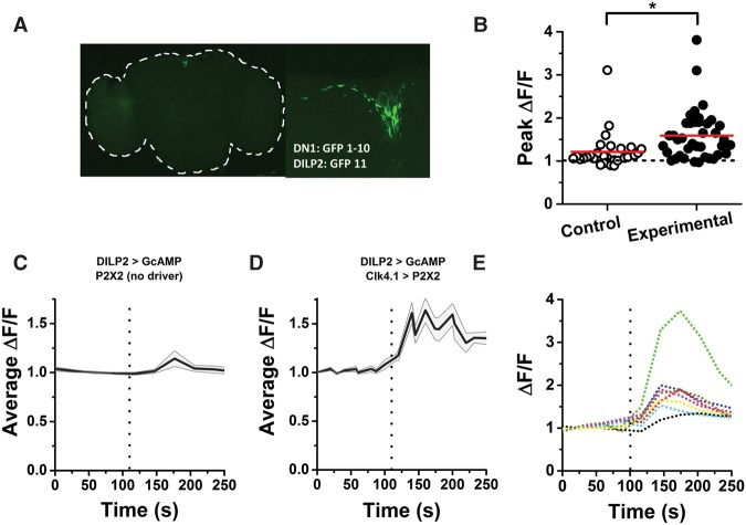
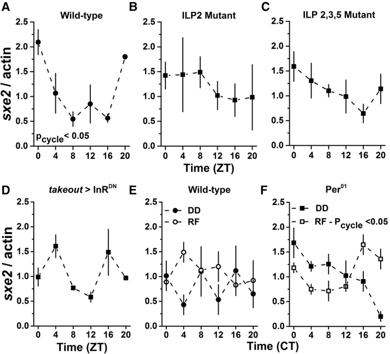
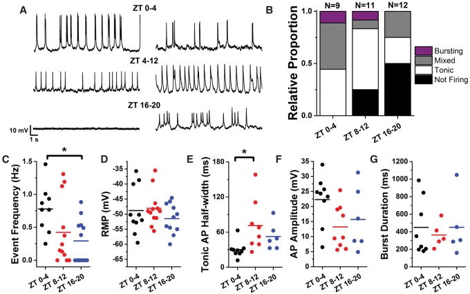
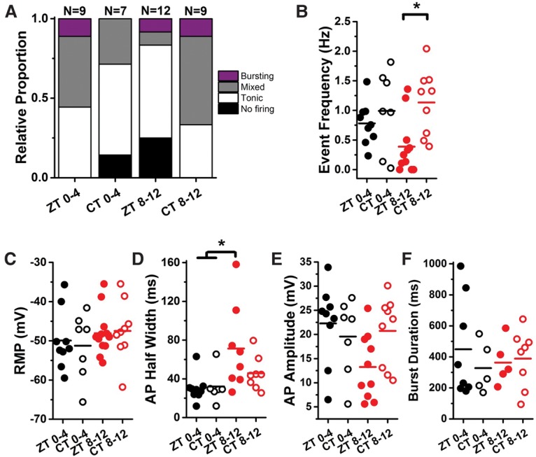
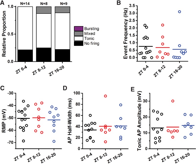
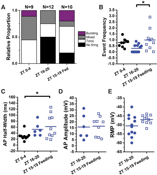
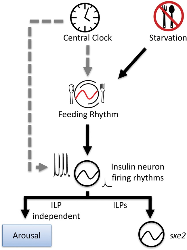
Circadian clocks regulate much of behavior and physiology, but the mechanisms by which they do so remain poorly understood. While cyclic gene expression is thought to underlie metabolic rhythms, little is known about cycles in cellular physiology. We found that Drosophila insulin-producing cells (IPCs), which are located in the pars intercerebralis and lack an autonomous circadian clock, are functionally connected to the central circadian clock circuit via DN1 neurons. Insulin mediates circadian output by regulating the rhythmic expression of a metabolic gene (sxe2) in the fat body. Patch clamp electrophysiology reveals that IPCs display circadian clock-regulated daily rhythms in firing event frequency and bursting proportion under light:dark conditions. The activity of IPCs and the rhythmic expression of sxe2 are additionally regulated by feeding, as demonstrated by night feeding-induced changes in IPC firing characteristics and sxe2 levels in the fat body. These findings indicate circuit-level regulation of metabolism by clock cells in Drosophila and support a role for the pars intercerebralis in integrating circadian control of behavior and physiology.

Keywords: Drosophila; circadian; clock; electrophysiology; insulin; metabolism.


Figures

Figure 1.
Figure 1.
IPCs are functionally connected to the circadian clock. (A) GRASP between DN1 neurons and IPCs (+; DILP2-Gal4/LexAOP-GFP11; Clk4.1-LexA/UAS-GFP1-10). Whole brain (left), outlined in white, and zoomed view of the PI region (right) showing the GFP signal in the PI and along the length of the projection from the DN1 to the PI. (B) Peak-normalized GCaMP6m fluorescence from individual IPCs after application of 2.5 mM ATP to stimulate control brains lacking P2X2 expression (control [open symbols]; mean ΔF/F = 1.2 ± 0.1) or DN1 cells expressing P2X2 receptors (experimental [filled symbols]; mean ΔF/F = 1.6 ± 0.1). Each point represents a single cell, and horizontal lines indicate the mean of all cells. The peak GCaMP excitation upon ATP application is significantly higher in the experimental group. P < 0.001 by Mann-Whitney test. Experimental fly genotype: +;DILP2-Gal4/UAS-mCherry.NLS;Clk4.1-LexA/LexAop-P2X2,UAS-GCaMP6m; control genotype: +;DILP2-Gal4/UAS-mCherry.NLS;+/LexAop-P2X2,UAS-GCaMP6m. (C) Averaged time course of GCaMP fluorescence in control flies. ATP was applied at the time indicated by the dashed line. Shading shows SEM. n = 29 cells from nine brains (five male and four female). (D) Averaged time course of GCaMP fluorescence in experimental flies as in C. n = 39 cells from eight brains (four male and four female). (E) Normalized time course of GCaMP6m fluorescence from eight IPCs in the same experimental brain shows the heterogeneity of the response to ATP applied at the time indicated by the dashed line.
Figure 2.
Figure 2.
sxe2 transcript rhythms in the fat body are regulated by insulin and feeding in the absence of a clock. (A) Wild-type (Iso31) flies displayed rhythmic sxe2 transcript expression in the fat body (P = 0.047) as assessed by JTK_Cycle analysis of quantitative RT–PCR (qRT–PCR) data. (B,C) Loss of DILP2 (B) or DILP2, DILP3, and DILP5 (C) resulted in loss of sxe2 transcript rhythm. P = 1.0 and P = 0.42, respectively. (D) Expression of a dominant-negative insulin receptor in the fat body (takeout-Gal4 > UAS-InRDN) resulted in a loss of sxe2 transcript rhythm. P = 0.88. (E) On the fourth day in DD, wild-type flies do not maintain rhythmic sxe2 transcript expression (filled symbols). P = 1.00. Restricted feeding (RF; open symbols) from circadian time (CT) 9–15 does not restore rhythmic sxe2 transcript expression. P = 0.11. (F) On the fourth day in DD, period mutant (per01) flies do not show rhythmic sxe2 transcript expression (filled symbols). P = 0.35. Restricted feeding (RF; open symbols) from CT 9–15 restores a circadian rhythm with a peak at CT 16. P = 2.0 × 10⁻⁴. n = 3 biological replicates per time point for AC, E, and F. n = 2 biological replicates per time point for D.
Figure 3.
Figure 3.
IPCs display circadian patterns of event frequency and morphology. (A) Fifteen-second representative traces from whole-cell patch clamp of GFP-labeled DILP2+ neurons from acutely dissected female Drosophila brains. Flies were entrained to a 12:12 LD cycle and sacrificed and recorded within the time window indicated. (B) Relative proportions of firing phenotypes from IPCs at different windows of the circadian day. (C) Event frequency of tonic and bursting events for three circadian time windows. Each point represents a single cell, and horizontal lines indicate the mean of all cells. Event frequency in the morning (ZT 0–4) is significantly different from night (ZT 16–20) by one-way ANOVA. P < 0.05. (D) Resting membrane potential for three circadian time windows. There was no time of day difference detected by one-way ANOVA. (E) Mean half-width of tonic action potentials for three circadian time windows. Tonic action potential (AP) half-width is significantly larger in the evening (ZT 8–12) than in the morning (ZT 0–4) by one-way ANOVA. P < 0.05. (F) Mean action potential amplitude for three circadian time windows. There was no time of day difference detected by one-way ANOVA. (G) Mean burst duration for three circadian time windows. There was no time of day difference detected by one-way ANOVA.
Figure 4.
Figure 4.
IPC electrophysiological rhythms are not maintained in DD. (A) Relative proportions of firing phenotypes from IPCs at different windows of the circadian day when entrained to a light cycle (ZT 0–4 and ZT 8–12) (data replotted from Fig. 1) compared with flies maintained in DD for 18–22 h prior to recording. CT 0–4, n = 7; CT 8–12, n = 9. (B) Event frequency was not different between ZT 0–4 and CT 0–4; however, we observed significantly higher event frequency at CT 8–12 compared with ZT 8–12. (C) Resting membrane potential was constant across all conditions. (D) Mean half-width of tonic action potentials was nearly identical between ZT 0–4 and CT 0–4 and also between ZT 8–12 and CT 8–12. Although the trend toward longer action potentials was maintained at CT 8–12, the half-width difference was not significant between CT 8–12 and morning time points. (E,F) Mean action potential amplitude (E) and mean burst duration (F) were similar across all conditions.
Figure 5.
Figure 5.
Ablation of the molecular clock results in a loss of electrophysiological rhythm. (A) Relative proportions of firing phenotypes from IPCs in per01 flies in the morning (ZT 0–4), evening (ZT 8–12), and night (ZT 16–20) showed nearly identical proportions of firing versus nonfiring cells and a loss of cells displaying burst firing events. (BE) Event frequency, resting membrane potential (RMP), tonic action potential (AP) half-width, and action potential (AP) amplitude from per01 flies at ZT 0–4 (black), ZT 8–12 (red), and ZT 16–20 (blue). Each point represents a single cell, and horizontal lines indicate the mean. There were no significant differences between time points for any parameter.
Figure 6.
Figure 6.
Restricting feeding during the night period partially restores “morning-like” firing properties. (A) Relative proportions of firing phenotypes from ZT 0–4 and ZT 16–20 (data replotted from Fig. 1) compared with flies starved for 18–22 h, fed for 2 h from ZT 12–14, and sacrificed for recording from ZT 15–19. (B) The event frequency for flies fed at night (open blue circles) is similar to the morning event frequency (black) and significantly higher than for control flies from the same time window fed ad lib (filled blue circles). (C) The mean tonic action potential half-width for flies fed at night (open blue circles) still resembles the night phenotype (filled blue circles) and is significantly different from the morning time window. (D,E) The mean action potential amplitude (D) and resting membrane potential (E) for flies fed ad lib versus flies with night-restricted feeding are not significantly different.
Figure 7.
Figure 7.
Model for dual modulation of IPCs by the circadian clock and feeding. Inputs from the brain clock can modulate IPC activity both directly via inputs from DN1 neurons and indirectly via clock control of feeding rhythms. Starvation can alter feeding rhythms, leading to altered IPC activity. IPC firing rhythms control insulin-dependent and insulin-independent downstream processes, which include metabolic transcript rhythms in the fat body and behavioral arousal.

References

    1. Alic N, Andrews TD, Giannakou ME, Papatheodorou I, Slack C, Hoddinott MP, Cochemé HM, Schuster EF, Thornton JM, Partridge L. 2011. Genome-wide dFOXO targets and topology of the transcriptomic response to stress and insulin signalling. Mol Syst Biol 7: 502. - PMC - PubMed
    1. Allada R, Chung BY. 2010. Circadian organization of behavior and physiology in Drosophila. Annu Rev Physiol 72: 605–624. - PMC - PubMed
    1. Arrese EL, Soulages JL. 2010. Insect fat body: energy, metabolism, and regulation. Annu Rev Entomol 55: 207–225. - PMC - PubMed
    1. Bai H, Kang P, Hernandez AM, Tatar M. 2013. Activin signaling targeted by insulin/dFOXO regulates aging and muscle proteostasis in Drosophila. PLoS Genet 9: e1003941. - PMC - PubMed
    1. Baslan T, Hicks J. 2014. Single cell sequencing approaches for complex biological systems. Curr Opin Genet Dev 26: 59–65. - PubMed

Publication types