. 2016 Dec 15;24(1):35-42.
doi: 10.1101/lm.043539.116. Print 2017 Jan.

Vagus nerve stimulation reduces cocaine seeking and alters plasticity in the extinction network

Affiliations

Vagus nerve stimulation reduces cocaine seeking and alters plasticity in the extinction network

Jessica E Childs et al. Learn Mem. .

Abstract

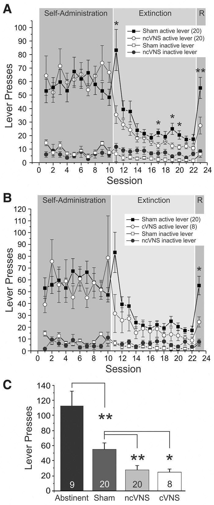
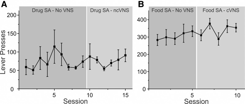
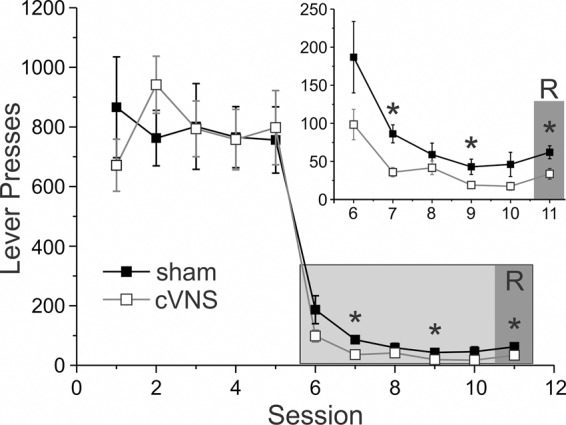
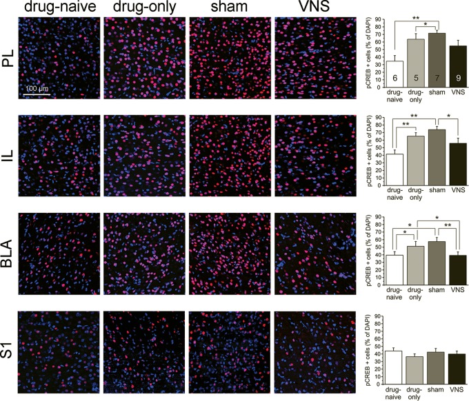
Drugs of abuse cause changes in the prefrontal cortex (PFC) and associated regions that impair inhibitory control over drug-seeking. Breaking the contingencies between drug-associated cues and the delivery of the reward during extinction learning reduces rates of relapse. Here we used vagus nerve stimulation (VNS) to induce targeted synaptic plasticity to facilitate extinction of appetitive behaviors and to reduce relapse. Rats self-administered cocaine and were given VNS during extinction. Relapse to drug-seeking was assessed in a cued reinstatement session. We used immunohistochemistry to measure changes in the expression of the phosphorylated transcription factor cAMP response-element binding protein (pCREB) in the PFC and the basolateral amygdala (BLA), which regulate cue learning and extinction. In vivo recordings of evoked field potentials measured drug- and VNS-induced changes in metaplasticity in the pathway from the PFC to the BLA. VNS-treated rats showed improved rates of extinction and reduced reinstatement. Following reinstatement, pCREB levels were reduced in the IL and BLA of VNS-treated rats. Evoked responses in the BLA were greatly reduced in VNS-treated rats, and these rats were also resistant to the induction of LTD. Taken together, these results show that VNS facilitates extinction and reduces reinstatement. Changes in the pathway between the PFC and the amygdala may contribute to these beneficial effects.


Figures

Figure 1.
Figure 1.
Vagus nerve stimulation (VNS) facilitates extinction from cocaine seeking and reduces cue-induced reinstatement. Rats were trained to self-administer cocaine and then underwent extinction training for 12 d while either receiving VNS (circles) or sham-stimulation (squares) before drug-seeking was reinstated by presentation of the previously drug-paired cues during a reinstatement session (R). (A) Comparison of sham-stimulated rats and rats receiving noncontingent VNS (ncVNS; see Materials and Methods for details of the stimulation parameters). (B) Comparison of sham-stimulated rats and rats receiving contingent VNS delivered with each lever press during extinction (cVNS; see Materials and Methods for details of the stimulation parameters). Rats receiving either ncVNS or cVNS showed reduced active lever presses during extinction and reinstatement. Inactive lever presses are shown as open circles. (C) Number of active lever presses during the reinstatement session for abstinent, sham-stimulated, and VNS treated rats. P values are (*) <0.05 and (**) <0.001.
Figure 2.
Figure 2.
Vagus nerve stimulation does not alter ongoing appetitive behavior. Rats were trained to self-administer cocaine (A) or food pellets (B). After a stable rate of response was reached, rats were given VNS during self-administration sessions to see whether this changes response patterns. Rats that self-administered cocaine received noncontingent VNS, while rats that self-administered food received contingent VNS with each lever press. Responses at the active lever during self-administration were not altered by VNS.
Figure 3.
Figure 3.
Vagus nerve stimulation facilitates extinction from food seeking. Rats were trained to self-administer food pellets for 5 d and then underwent extinction training for 5 d while they received either VNS (open squares) or sham-stimulation (black squares). Food seeking was reinstated by presentation of the previously food-paired cues during a reinstatement session (R). Compared with sham-stimulated rats VNS rats made fewer responses at the active lever during extinction and during the cue-induced reinstatement session. However, rats in both groups showed only small, nonsignificant increases in responses during cue-induced reinstatement when compared with the last day of extinction . P values are (*) <0.05. Inset shows a detail of the extinction and reinstatement days.
Figure 4.
Figure 4.
Vagus nerve stimulation reduces pCREB expression in infralimbic cortex and basolateral amygdala following reinstatement of drug-seeking. Photomicrographs (20× magnification) of phosphorylated CREB expression (red) in the prelimbic (PL, top row) and infralimbic (IL, second row) prefrontal cortex, the basolateral amygdala (BLA, third row), as well as the primary somatosensory cortex (S1, bottom row) in four treatment groups (drug-naïve, drug-only, sham-stimulated, and noncontingent VNS). Bargraphs on the right summarize the data for each row (PL, IL, BLA, S1). pCREB expression is shown as the percentage of cell bodies that showed pCREB immunoreactivity among all DAPI-stained cells. Rats trained to self-administer cocaine (drug-only, light gray bars) and sham-stimulated rats (dark gray bars) showed increased pCREB expression in the PL, IL, and BLA compared with drug-naïve rats (white bars). VNS treatment (black bars) given during extinction reduced pCREB expression in the IL and BLA compared with sham-stimulated rats to similar levels as seen in drug-naïve rats. P values are (*) <0.05 and (**) <0.001.
Figure 5.
Figure 5.
VNS treatment alters the responsiveness of the basolateral amygdala to stimulation of the infralimbic cortex. (A) Diagram of the stimulation and recording sites in the infralimbic (IL) PFC and the BLA. (B) Single-pulse stimulation targeted to the IL elicited negative field potentials in the BLA that peaked after 20–25 msec. Representative traces (average of six consecutive sweeps) of an input–output curve from a drug-naive rat. (C) Input–output curves from rats in five treatment groups (drug-naïve, black squares; drug-only, triangles; abstinent, gray circles; sham-stimulated, white squares; non-contingent VNS, black circle). VNS rats showed reduced amplitudes of the evoked local field potential compared with drug-naïve and abstinent rats. (D) Low-frequency (1 Hz) stimulation applied at baseline stimulation intensity (40% of the stimulation intensity that evoked a maximum field response) induced LTD in drug-naïve and sham-stimulated rats. All other groups show no significant change from baseline following low-frequency stimulation, despite differences in the range of field potential amplitudes that could be evoked under baseline conditions (C).

References

    1. Aihua L, Lu S, Liping L, Xiuru W, Hua L, Yuping W. 2014. A controlled trial of transcutaneous vagus nerve stimulation for the treatment of pharmacoresistant epilepsy. Epilepsy Behav 39: 105–110. - PubMed
    1. Chase HM, Sterman MB, Clemente CD. 1966. Cortical and subcortical patters of response to afferent vagal stimulation. Exp Neurol 16: 36–49. - PubMed
    1. Childs JE, Alvaraz-Dieppa A, McIntyre C, Kroener S. 2015. Vagus nerve stimulation as a tool to induce plasticity in pathways relevant for extinction learning. J Vis Exp 102: e53032. - PMC - PubMed
    1. Clark KB, Krahl SE, Smith DC, Jensen RA. 1995. Post-training unilateral vagal stimulation enhances retention performance in the rat. Neurobiol Learn Mem 63: 213–216. - PubMed
    1. Conklin CA, Tiffany ST. 2002. Applying extinction research and theory to cue-exposure addiction treatments. Addiction 97: 155–167. - PubMed

MeSH terms