. 2017 Apr;65(4):1249-1266.
doi: 10.1002/hep.28964. Epub 2017 Jan 31.

Prohibitin 1 suppresses liver cancer tumorigenesis in mice and human hepatocellular and cholangiocarcinoma cells

Affiliations

Prohibitin 1 suppresses liver cancer tumorigenesis in mice and human hepatocellular and cholangiocarcinoma cells

Wei Fan et al. Hepatology. 2017 Apr.

Erratum in

  • Correction.
    [No authors listed] [No authors listed] Hepatology. 2017 Nov;66(5):1708. doi: 10.1002/hep.29550. Hepatology. 2017. PMID: 29053195 No abstract available.

Abstract

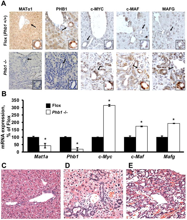
Prohibitin 1 (PHB1) is best known as a mitochondrial chaperone, and its role in cancer is conflicting. Mice lacking methionine adenosyltransferase α1 (MATα1) have lower PHB1 expression, and we reported that c-MYC interacts directly with both proteins. Furthermore, c-MYC and MATα1 exert opposing effects on liver cancer growth, prompting us to examine the interplay between PHB1, MATα1, and c-MYC and PHB1's role in liver tumorigenesis. We found that PHB1 is highly expressed in normal hepatocytes and bile duct epithelial cells and down-regulated in most human hepatocellular carcinoma (HCC) and cholangiocarcinoma (CCA). In HCC and CCA cells, PHB1 expression correlates inversely with growth. PHB1 and MAT1A positively regulate each other's expression, whereas PHB1 negatively regulates the expression of c-MYC, MAFG, and c-MAF. Both PHB1 and MATα1 heterodimerize with MAX, bind to the E-box element, and repress E-box promoter activity. PHB1 promoter contains a repressive E-box element and is occupied mainly by MAX, MNT, and MATα1 in nonmalignant cholangiocytes and noncancerous tissues that switched to c-MYC, c-MAF, and MAFG in cancer cells and human HCC/CCA. All 8-month-old liver-specific Phb1 knockout mice developed HCC, and one developed CCA. Five-month-old Phb1 heterozygotes, but not Phb1 flox mice, developed aberrant bile duct proliferation; and one developed CCA 3.5 months after left and median bile duct ligation. Phb1 heterozygotes had a more profound fall in the expression of glutathione synthetic enzymes and higher hepatic oxidative stress following left and median bile duct ligation.

Conclusion: We have identified that PHB1, down-regulated in most human HCC and CCA, heterodimerizes with MAX to repress the E-box and positively regulates MAT1A while suppressing c-MYC, MAFG, and c-MAF expression; in mice, reduced PHB1 expression predisposes to the development of cholestasis-induced CCA. (Hepatology 2017;65:1249-1266).


Figures

Figure 1
Figure 1. PHB1 interacts with MATα1, c-MYC, MAFG and c-MAF
(A) The liver protein lysates from a 3-month old Phb1 flox male mouse liver (12) were subjected to immunoprecipitation (IP) using anti-PHB1 or nonspecific IgG, followed by Western blotting for MAFG, MATα1, c-MYC, c-MAF, PHB1, and GAPDH (as negative control). (B) In vitro pull down assay using immobilized recombinant PHB1 or MATα1 demonstrates direct interaction between PHB1 and MATα1. (C) In vitro pull down assay using immobilized recombinant PHB1 or MAFG demonstrates direct interaction between PHB1 and MAFG. Results represent a total of at least 3 independent experiments.
Figure 2
Figure 2. PHB1 suppresses growth in HCC and CCA cells, is down-regulated in human HCC and CCA
Effect of varying PHB1 expression on growth of two HCC (HepG2 and Hep3B) and two CCA (KMCH and Huh28) cell lines was assessed using BrdU as described in Experimental Procedures. (A) Overexpression of PHB1 suppressed BrdU incorporation, similar to overexpression of MAT1A or knocking down c-MAF, MAFG or c-MYC. In contrast, overexpression of c-MAF, MAFG, and c-MYC all increased BrdU incorporation, similar to knocking down MAT1A or PHB1. *p < 0.05 versus respective controls. (B) Immunohistochemical (IHC) analysis of PHB1, MATα1, c-MYC, MAFG and c-MAF in paraffin-embedded specimens from human HCC and CCA. In normal liver, PHB1 is highly expressed in hepatocytes and bile duct epithelial cells but it is down-regulated in CCA. PHB1 and MATα1 are highly expressed in non-tumorous livers and down-regulated in HCC. In contrast, c-MYC, MAFG and c-MAF have low expression in non-tumorous livers and are up-regulated in HCC. Arrows point to the small bile duct (insets at the bottom right of the IHC image). Original magnification, ×200. (C) PHB1 mRNA levels in 4 independent HCC microarray from GEO database as compared to adjacent non-tumor tissue. (D) PHB1 mRNA levels from the only GEO database that contains information on CCA (GSE15765) show lower expression in CCA as compared to HCC. The database normalized PHB1 expression by the RMA (robust multi-array average) method (31).
Figure 3
Figure 3. Interplay among PHB1, MAT1A, c-MYC, MAFG and c-MAF
PHB1 expression was varied by overexpression (A) or knockdown (B) in HepG2, Hep3B, KMCH and Huh28 cells and the expression of MAT1A, c-MYC, MAFG and c-MAF was measured as described in Experimental Procedures. (C) PHB1 expression was measured in the same four cell lines after varying MAT1A, c-MYC, MAFG and c-MAF expression. Results are expressed as mean ± SEM from at least 3 independent experiments, *p < 0.05 versus respective controls. Protein expression was measured in HepG2 and Huh28 cells (results shown are for HepG2 cells, with similar results found in Huh28 cells).
Figure 4
Figure 4. PHB1 and MATα1 heterodimerize with MAX, bind to the E-box and repress E-box driven promoter activity
(A) EMSA analysis of a multimerized human PHB1 E-box element (X3) using recombinant PHB1, MATα1, c-MYC and MAX proteins in different combinations. PHB1 cannot bind to E-box alone or with c-MYC but can bind to E-box in the presence of MAX, similar to MATα1. (B) Promoter activity of a consensus E-box-driven luciferase reporter construct and its mutant after overexpression or knockdown of PHB1 in two HCC and two CCA cell lines. *p< 0.05 vs respective controls. Results represent a total of at least 3 independent experiments done in duplicate.
Figure 5
Figure 5. Occupancy of PHB1 promoter E-box and its regulation by MATα1, c-MYC, MAFG and c-MAF
(A) H69 (benign bile duct epithelial cells), Huh28 and KMCH (CCA cells) and HepG2 (HCC cells) were subjected to ChIP analysis with MAX followed by Seq-ChIP with c-MYC, MAFG, c-MAF, MNT, MATα1, or PHB1 spanning the E-box containing human PHB1 promoter (shown on top) as described in Experimental Procedures. (B) ChIP was done using human HCC, CCA and their adjacent non-tumorous tissues with MAX followed by Seq-ChIP with c-MYC, MAFG, c-MAF, MNT, MATα1, or PHB1 spanning the E-box containing human PHB1 promoter as in (A). Representatives from three pairs of HCC and CCA are shown. (C) Effect of overexpressing MAT1A, c-MYC, MAFG or c-MAF on human PHB1 promoter (wild type or E-box mutant) was examined in Huh28 and HepG2 cells. *p< 0.05 vs control (empty vector). Results represent a total of 3 independent experiments done in duplicate.
Figure 6
Figure 6. Liver-specific Phb1 KO mice have altered MAT1A, c-MYC, MAFG and c-MAF expression, develop HCC and CCA
Expression of PHB1, MATα1, c-MYC, MAFG and c-MAF was measured using (A) IHC (arrows point to the bile duct in the insets at the bottom right of each IHC image) and (B) real-time PCR in liver-specific Phb1 KO mice livers (n=3 for each group). Comparison of liver histology on hematoxylin and eosin (H&E) between (C) 8-months old flox controls (normal histology) and liver-specific Phb1 KO mice (all 8 mice have HCC, not shown) show (D) atypical hyperplasia of bile ducts in 6 out of 8 mice, and (E) CCA in 1 out of 8. Original magnification, X200 for all.
Figure 7
Figure 7. Liver-specific Phb1 heterozygotes are sensitized to develop LMBDL-induced CCA
5-month old liver-specific Phb1 heterozygotes and flox controls (n=8 each group) were subjected to LMBDL as described in Experimental Procedures and followed for 3.5 months. (A) Flox control (left panel); heterozygous control (middle panel), showing proliferation of bile duct epithelial cells in 5/8 at baseline; flox LMBDL control (right panel), where bile duct proliferation was found in portal tract in 8/8; (B) heterozygous LMBDL mice exhibited aberrant bile duct proliferation (left panel) in 8/8; CCA (middle panel) was found in 1/8 heterozygotes after LMBDL with vascular invasion (right panel, arrow), (C) invasion to adjacent tissue (left panel, arrows), pancreas (middle panel) and lung metastasis (right panel, arrow). Representative H&E are shown, original magnification, X200 for all except X400 for F and G. (D) Changes in Phb1, Mat1a, c-Myc, Mafg and c-Maf expression after LMBDL, expressed as % of Phb1 flox control. Real-time PCR measured mRNA levels of these genes from n=3 in each group. *p< 0.05 vs Flox, †p < 0.05 vs Phb1 flox + LMBDL. (E) Western blotting showed comparable changes in PHB1 protein levels.
Figure 8
Figure 8. PHB1 is a tumor suppressor in SAMe-D cells
(A) PHB1 expression is lower in SAMe-D cells, which are derived from HCC of a Mat1a KO mouse. (B) Effect of PHB1 overexpression on expression of c-MYC, c-MAF, and MAFG in SAMe-D cells. (C) Anchorage-independent growth in SAMe-D cells. SAMe-D cells were stably transfected with PHB1 overexpression vector as described in Experimental Procedures and anchorage-independent growth was measured. Overexpression of PHB1 had a profound inhibitory effect on anchorage-independent growth.

References

    1. Theiss AL, Sitaraman SV. The role and therapeutic potential of prohibitin in disease. Biochim Biophys Acta. 2011;1813:1137–1143. - PMC - PubMed
    1. Nijtmans LGJ, Sanz MA, Grivell LA, Coates PJ. The mitochondrial PHB complex: roles in mitochondrial respiratory complex assembly, ageing and degenerative disease. Cell Mol Life Sci. 2002;59:143–155. - PMC - PubMed
    1. Wang S, Fusaro G, Padmanabhan J, Chellappan SP. Prohibitin co-localizes with Rb in the nucleus and recruits N-CoR and HDAC1 for transcriptional repression. Oncogene. 2002;21:8388–8396. - PubMed
    1. Choi D, Lee SJ, Kim IH, Kang S. Prohibitin interacts with RNF2 and regulates E2F1 function via dual pathways. Oncogene. 2008;27:1716–1725. - PubMed
    1. Fusaro G, Dasgupta P, Rastogi S, Joshi B, Chellappan S. Prohibitin induces the transcriptional activity of p53 and is exported from the nucleus upon apoptotic signaling. J Biol Chem. 2003;278:47853–47861. - PubMed

MeSH terms