

Xenogeneic Decellularized Scaffold: A Novel Platform for Ovary Regeneration
- PMID: 27981878
- PMCID: PMC5315000
- DOI: 10.1089/ten.TEC.2016.0410
Xenogeneic Decellularized Scaffold: A Novel Platform for Ovary Regeneration
Erratum in
-
Correction to: Xenogeneic Decellularized Scaffold: A Novel Platform for Ovary Regeneration doi: 10.1089/ten.tec.2016.0410.Tissue Eng Part C Methods. 2025 Apr;31(4):164-165. doi: 10.1089/ten.tec.2016.0410.correx. Tissue Eng Part C Methods. 2025. PMID: 40249237 Free PMC article. No abstract available.
Abstract
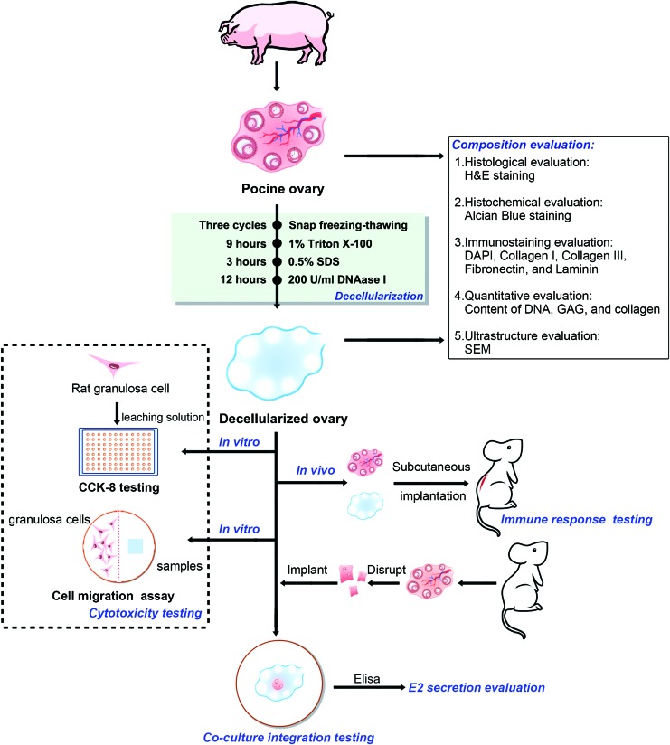
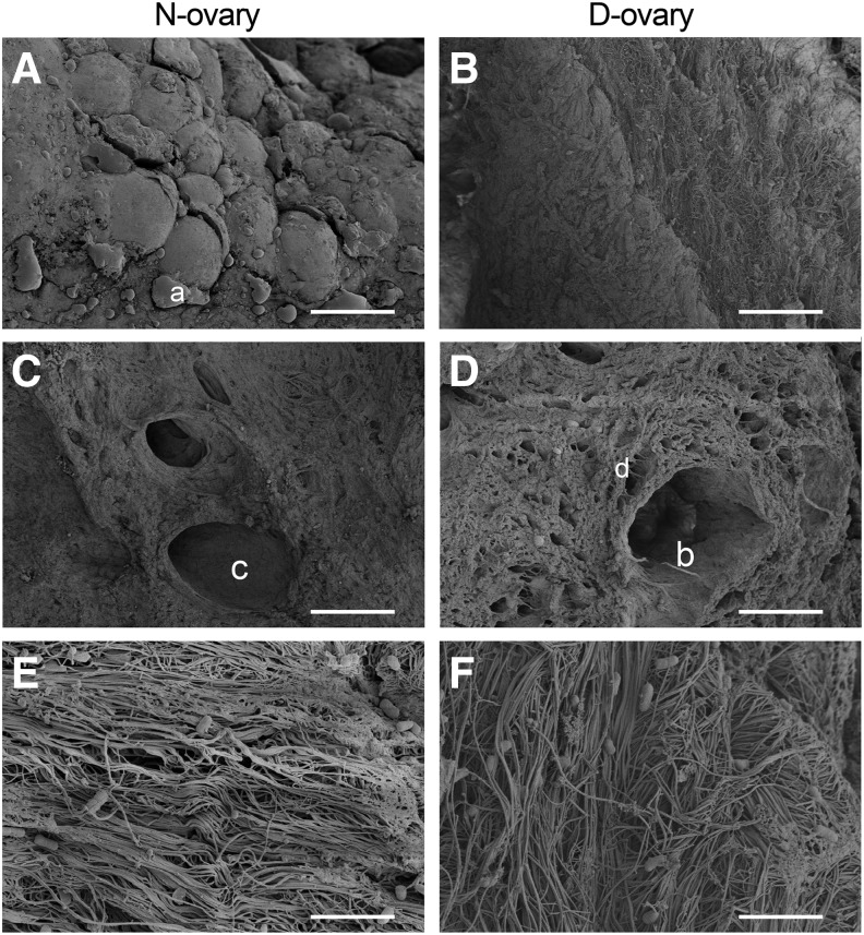
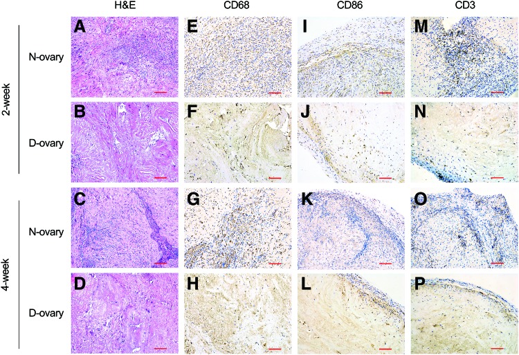
Women younger than 40 years may face early menopause because of premature ovarian failure (POF). The cause of POF can be idiopathic or iatrogenic, especially the cancer-induced oophorectomy and chemo- or radiation therapy. The current treatments, including hormone replacement therapy (HRT) and cryopreservation techniques, have increased risk of ovarian cancer and may reintroduce malignant cells after autografting. Decellularization technique has been regarded as a novel regenerative medicine strategy for organ replacement, wherein the living cells of an organ are removed, leaving the extracellular matrix (ECM) for cellular seeding. This study aimed to produce a xenogeneic decellularized ovary (D-ovary) scaffold as a platform for ovary regeneration and transplantation. We have developed a novel decellularization protocol for porcine ovary by treatment with physical, chemical, and enzymatic methods. Using hematoxylin and eosin (H&E) staining, DAPI staining, scanning electron microscopy (SEM), and quantitative analysis, this approach proved effective in removing cellular components and preserving ECM. Furthermore, the results of biological safety evaluation demonstrated that the D-ovary tissues were noncytotoxic for rat ovarian cells in vitro and caused only a minimal immunogenic response in vivo. In addition, the D-ovary tissues successfully supported rat granulosa cell penetration ex vivo and showed an improvement in estradiol (E2) hormone secretion.
Keywords: bioengineering in organ transplantation; decellularization; extracellular matrix; premature ovarian failure; regenerative medicine.
Conflict of interest statement
No competing financial interests exist.
Figures


References
-
- Luborsky J.L., Meyer P., Sowers M.F., Gold E.B., and Santoro N. Premature menopause in a multi-ethnic population study of the menopause transition. Hum Reprod 18, 199, 2003 - PubMed
-
- Laissue P. Aetiological coding sequence variants in non-syndromic premature ovarian failure: from genetic linkage analysis to next generation sequencing. Mol Cell Endocrinol 411, 243, 2015 - PubMed
-
- Osterlund M.K., Witt M.R., and Gustafsson J.A. Estrogen action in mood and neurodegenerative disorders: estrogenic compounds with selective properties-the next generation of therapeutics. Endocrine 28, 235, 2005 - PubMed
Publication types
MeSH terms
Substances
LinkOut - more resources
Full Text Sources
Other Literature Sources
