Review
. 2017 Jan;13(1):52-64.
doi: 10.1038/nrneurol.2016.185. Epub 2016 Dec 16.

Mechanisms of radiotherapy-associated cognitive disability in patients with brain tumours

Affiliations
Review

Mechanisms of radiotherapy-associated cognitive disability in patients with brain tumours

Milan T Makale et al. Nat Rev Neurol. 2017 Jan.

Abstract

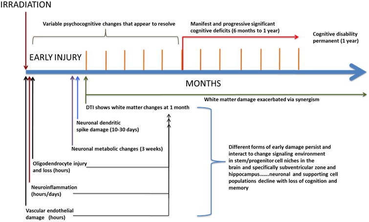
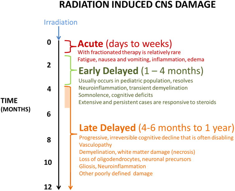
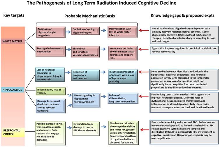
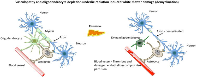
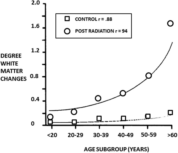
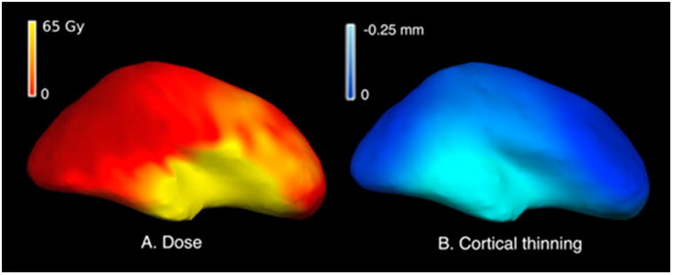
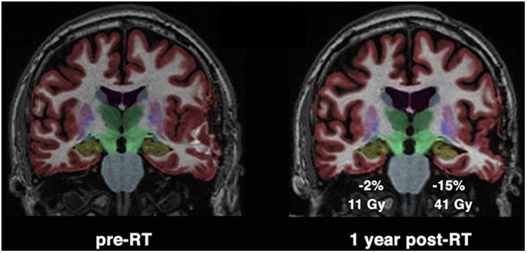
Standard treatment of primary and metastatic brain tumours includes high-dose megavoltage-range radiation to the cranial vault. About half of patients survive >6 months, and many attain long-term control or cure. However, 50-90% of survivors exhibit disabling cognitive dysfunction. The radiation-associated cognitive syndrome is poorly understood and has no effective prevention or long-term treatment. Attention has primarily focused on mechanisms of disability that appear at 6 months to 1 year after radiotherapy. However, recent studies show that CNS alterations and dysfunction develop much earlier following radiation exposure. This finding has prompted the hypothesis that subtle early forms of radiation-induced CNS damage could drive chronic pathophysiological processes that lead to permanent cognitive decline. This Review presents evidence of acute radiation-triggered CNS inflammation, injury to neuronal lineages, accessory cells and their progenitors, and loss of supporting structure integrity. Moreover, injury-related processes initiated soon after irradiation could synergistically alter the signalling microenvironment in progenitor cell niches in the brain and the hippocampus, which is a structure critical to memory and cognition. Progenitor cell niche degradation could cause progressive neuronal loss and cognitive disability. The concluding discussion addresses future directions and potential early treatments that might reverse degenerative processes before they can cause permanent cognitive disability.


Figures

Figure 1
Figure 1. Evidence for early radiation injury to the CNS
A proposed time course and scheme by which early damage becomes chronic and the interactions leading to permanent cognitive disability.
Figure 2
Figure 2
Manifestations and time course of radiation induced CNS injury and cognitive decline.
Figure 3
Figure 3. Primary radiation induced CNS abnormalities
Detailed scheme shows major proposed mechanisms contributing to radiation induced cognitive decline.
Figure 4
Figure 4. The presumed origins of white matter damage
White matter provides essential connectivity for cortical function. Oligodendrocytes establish and maintain myelin around white matter axons (left panel) and their destruction (right panel) results in a loss of myelin integrity. Damage to feeding microvessels (right panel) also compromises white matter but also has adverse effects on the perfusion of other key CNS elements such as astrocytes which provide metabolic and functional support to neurons.
Figure 5
Figure 5. The severity of radiation induced white matter damage increases with age
Graph of the average degree of supratentorial white matter changes versus age. Therapeutic and controls groups are included, the curves were fitted and correlation calculated using second-order polynomial regression. Taken from Tsuruda et al (AJR 149:165-171, 1987). Used with permission.
Figure 6
Figure 6. Cortical thinning according to dose at one year post-irradiation
Cortical surface representation of (A) radiation dose in Gy and (B) cortical thinning at 1 year observed in an example patient. Regions receiving a higher dose show a greater degree of cortical thinning at 1 year post radiation therapy.
Figure 7
Figure 7. Radiation dose-dependent hippocampal atrophy
Coronal projection of brain MRI from a glioma patient pre-radiotherapy (pre-RT) on left, and one year post-radiotherapy (post-RT) on right. Color overlay shows automated segmentation of hippocampus in yellow. Greater percent decrease in hippocampal volume was seen in the hippocampus with higher mean dose of 41 Gy.

References

    1. Chi A, Komaki R. Treatment of brain metastasis from lung cancer. Cancers (Basel) 2010;2:2100–2137. - PMC - PubMed
    1. Shi L, et al. Aging masks detection of radiation-induced brain injury. Brain Res. 2011;1385:307–316. - PMC - PubMed
    1. Greene-Schloesser D, Moore E, Robbins ME. Molecular pathways: radiation-induced cognitive impairment. Clin Cancer Res. 2013;19:2294–2300. - PMC - PubMed
    1. Moore ED, Kooshki M, Wheeler KT, Metheny-Barlow LJ, Robbins ME. Differential expression of Homer1a in the hippocampus and cortex likely plays a role in radiation-induced brain injury. Radiat Res. 2014;181:21–32. - PMC - PubMed
    1. Wu PH, et al. Radiation induces acute alterations in neuronal function. PLoS One. 2012;7:e37677. - PMC - PubMed

MeSH terms