. 2016 Dec 16:6:39297.
doi: 10.1038/srep39297.

Unraveling gene expression profiles in peripheral motor nerve from amyotrophic lateral sclerosis patients: insights into pathogenesis

Affiliations

Unraveling gene expression profiles in peripheral motor nerve from amyotrophic lateral sclerosis patients: insights into pathogenesis

Nilo Riva et al. Sci Rep. .

Abstract

The aim of the present study is to investigate the molecular pathways underlying amyotrophic lateral sclerosis (ALS) pathogenesis within the peripheral nervous system. We analyzed gene expression changes in human motor nerve diagnostic biopsies obtained from eight ALS patients and seven patients affected by motor neuropathy as controls. An integrated transcriptomics and system biology approach was employed. We identified alterations in the expression of 815 genes, with 529 up-regulated and 286 down-regulated in ALS patients. Up-regulated genes clustered around biological process involving RNA processing and protein metabolisms. We observed a significant enrichment of up-regulated small nucleolar RNA transcripts (p = 2.68*10-11) and genes related to endoplasmic reticulum unfolded protein response and chaperone activity. We found a significant down-regulation in ALS of genes related to the glutamate metabolism. Interestingly, a network analysis highlighted HDAC2, belonging to the histone deacetylase family, as the most interacting node. While so far gene expression studies in human ALS have been performed in postmortem tissues, here specimens were obtained from biopsy at an early phase of the disease, making these results new in the field of ALS research and therefore appealing for gene discovery studies.


Figures

Figure 1
Figure 1. Representative neuropathological cases.
Transverse semi-thin sections of biopsy of motor nerve from an ALS case (A and B) and motor neuropathy patient (C and D). In ALS patients, focal decreased density of myelinated nerve fibers (A and B: asterisk) is evident in a nerve fascicles. At higher magnification (B), axonal degeneration is evident (arrows). (C) Diffuse mild reduction of myelin nerve fibers is present in a representative section from patients with definite diagnosis of motor neuropahty. There are clusters of small myelinated fibers (D, arrows), indicating axonal regeneration. In addition small onion bulbs (D, arrowheads), indicating remyelination, are present. Bar: (A,C) 50 μm; (B,D) 10 μm.
Figure 2
Figure 2. Heatmap representing hierarchical clustering of the 15 samples (columns) and genes (rows) detected as differentially expressed according to two different thresholds.
(A) FDR < 0.01 and FC > 1.5 or FC < 0.66; (B) FDR < 0.05 and FC > 3 or FC < 0.33. The expression level of each gene has been standardized by subtracting the gene’s mean expression level and dividing by the standard deviation across all samples. This scaled expression value is plotted in red-green scale color, with red indicating higher expression and green lower expression in ALS patients.
Figure 3
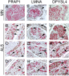
Figure 3. Immunohistochemistry localization of PRAP1, LMNA and DPYSL4 in MN and ALS motor nerves.
Double staining for MBP (red), to mark myelinated nerve fibers, and PRAP1 (AD), LMNA (EH) and DPYSL4 (IL) (brown) shows increase expression in ALS motor nerves. PRAP1 immunoreactivity was present in the endoneurium in numeorus cells (arrowheads, B,C) including cytoplasm of Schwann cells (arrows, B; MBP immunoreactivity identified the myelin sheath) and endothelial cells (arrow, D). LMNA shows immunoreactivity in the perineurium in both MN and ALS nerves (arrowhead, E,F); in ALS motor nerves, immunoreactivity was present in nuclei and cytoplasm of all endoneurial cells (arrowhead, G,H) and Schwann cells (arrows, F–H). In ALS motor nerves, DPYSL4 was expressed in perineurium (arrowhead, J) and in the cytoplams and nuclei of many cells (arrows in JL) including endothelial cells (arrowhead, K) and Schwann cells (arrows J,K). Bar: (A,E,I) 50 μm; (F–H,J–L) 50 μm.
Figure 4
Figure 4. Expression of NOP14 in the sciatic nerve of normal and SOD1G93A mice.
Tranverse sections from wild-type (AD), SOD1G93A mice at 90 days (diseases onset) (EH) and 120 (end-stage disease) (IL) double-stained for neurofilament (green), to recognize axons, and NOP14 (red). DAPI staining of nuclei (blue). Nop14 immunoreactivity was present in cell nuclei in wild-type nerves (arrowhead, merge in D); NOP14 shows an increase expression in SOD1G93A nerves in cell nuclei of the Schwann cells (arrowhead, merge in H and L); perineurial cells were also positive for NOP14 (merge in H and L, arrows). Bar: (A–L) 120 μm.
Figure 5
Figure 5. Interaction networks among differentially expressed genes as retrieved from STRING database at confidence score >0.7.
(A) Largest connected network, with subnetwork modules mined by ClusterONE algorithm as densely connected regions, highlighted in (BD). Nodes are color-coded according to values of log2 (fold-change), with red and green nodes representing up-regulated and down-regulated genes respectively, and color saturation prorportional to level of modulation. Node size is proportional to degree connectivity and border thickness is proportional to adjusted p-value of association.

References

    1. Ravits J. et al.. Deciphering amyotrophic lateral sclerosis: what phenotype, neuropathology and genetics are telling us about pathogenesis. Amyotroph Lateral Scler Frontotemporal Degener 14, 5–18 (2013). - PMC - PubMed
    1. Riva N., Agosta F., Lunetta C., Filippi M. & Quattrini A. Recent advances in amyotrophic lateral sclerosis. J Neurol 263, 1241–1254 (2016). - PMC - PubMed
    1. Renton A. E., Chio A. & Traynor B. J. State of play in amyotrophic lateral sclerosis genetics. Nat Neurosci 17, 17–23 (2014). - PMC - PubMed
    1. Cleveland D. W. & Rothstein J. D. From Charcot to Lou Gehrig: deciphering selective motor neuron death in ALS. Nat Rev Neurosci 2, 806–819 (2001). - PubMed
    1. Ilieva H., Polymenidou M. & Cleveland D. W. Non-cell autonomous toxicity in neurodegenerative disorders: ALS and beyond. J Cell Biol 187, 761–772 (2009). - PMC - PubMed

Publication types