

Differential analysis of genome-wide methylation and gene expression in mesenchymal stem cells of patients with fractures and osteoarthritis
- PMID: 27982725
- PMCID: PMC5330439
- DOI: 10.1080/15592294.2016.1271854
Differential analysis of genome-wide methylation and gene expression in mesenchymal stem cells of patients with fractures and osteoarthritis
Abstract
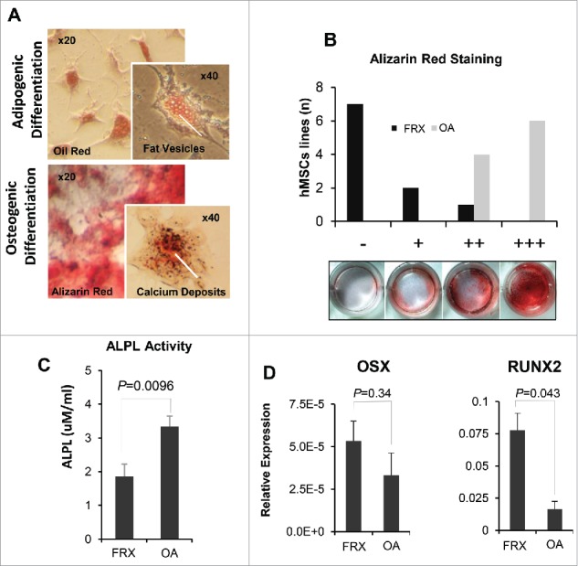
Insufficient activity of the bone-forming osteoblasts leads to low bone mass and predisposes to fragility fractures. The functional capacity of human mesenchymal stem cells (hMSCs), the precursors of osteoblasts, may be compromised in elderly individuals, in relation with the epigenetic changes associated with aging. However, the role of hMSCs in the pathogenesis of osteoporosis is still unclear. Therefore, we aimed to characterize the genome-wide methylation and gene expression signatures and the differentiation capacity of hMSCs from patients with hip fractures. We obtained hMSCs from the femoral heads of women undergoing hip replacement due to hip fractures and controls with hip osteoarthritis. DNA methylation was explored with the Infinium 450K bead array. Transcriptome analysis was done by RNA sequencing. The genomic analyses revealed that most differentially methylated loci were situated in genomic regions with enhancer activity, distant from gene bodies and promoters. These regions were associated with differentially expressed genes enriched in pathways related to hMSC growth and osteoblast differentiation. hMSCs from patients with fractures showed enhanced proliferation and upregulation of the osteogenic drivers RUNX2/OSX. Also, they showed some signs of accelerated methylation aging. When cultured in osteogenic medium, hMSCs from patients with fractures showed an impaired differentiation capacity, with reduced alkaline phosphatase activity and poor accumulation of a mineralized matrix. Our results point to 2 areas of potential interest for discovering new therapeutic targets for low bone mass disorders and bone regeneration: the mechanisms stimulating MSCs proliferation after fracture and those impairing their terminal differentiation.
Keywords: Epigenetics; osteoarthritis; osteoporosis; stem cells; transcription factors.
Figures


References
-
- Liu H, Xia X, Li B. Mesenchymal stem cell aging: Mechanisms and influences on skeletal and non-skeletal tissues. Exp Biol Med (Maywood) 2015; 240:1099-106; PMID:26088863; http://dx.doi.org/10.1177/1535370215591828 - DOI - PMC - PubMed
-
- Donoso O, Pino AM, Seitz G, Osses N, Rodríguez JP. Osteoporosis-associated alteration in the signalling status of BMP-2 in human MSCs under adipogenic conditions. J Cell Biochem 2015; 116:1267-77; PMID:25640452; http://dx.doi.org/10.1002/jcb.25082 - DOI - PubMed
-
- Haasters F, Docheva D, Gassner C, Popov C, Böcker W, Mutschler W, Schieker M, Prall WC. Mesenchymal stem cells from osteoporotic patients reveal reduced migration and invasion upon stimulation with BMP-2 or BMP-7. Biochem Biophys Res Commun 2014; 452:118-23; PMID:25152406; http://dx.doi.org/10.1016/j.bbrc.2014.08.055 - DOI - PubMed
-
- Liu W, Qi M, Konermann A, Zhang L, Jin F, Jin Y. The p53/miR-17/Smurf1 pathway mediates skeletal deformities in an age-related model via inhibiting the function of mesenchymal stem cells. Aging (Albany NY) 2015; 7:205-18; PMID:25855145; http://dx.doi.org/10.18632/aging.100728 - DOI - PMC - PubMed
-
- Prall WC, Haasters F, J Heggebö, Polzer H, Schwarz C, Gassner C, Grote S, Anz D, Jäger M, Mutschler W, et al.. Mesenchymal stem cells from osteoporotic patients feature impaired signal transduction but sustained osteoinduction in response to BMP-2 stimulation. Biochem Biophys Res Commun 2013; 440:617-22; PMID:24099772; http://dx.doi.org/10.1016/j.bbrc.2013.09.114 - DOI - PubMed
MeSH terms
LinkOut - more resources
Full Text Sources
Other Literature Sources
Medical