Review
. 2016 Dec 14;17(12):2095.
doi: 10.3390/ijms17122095.

Molecular Mechanisms and Translational Therapies for Human Epidermal Receptor 2 Positive Breast Cancer

Affiliations
Review

Molecular Mechanisms and Translational Therapies for Human Epidermal Receptor 2 Positive Breast Cancer

Quanxia Lv et al. Int J Mol Sci. .

Abstract

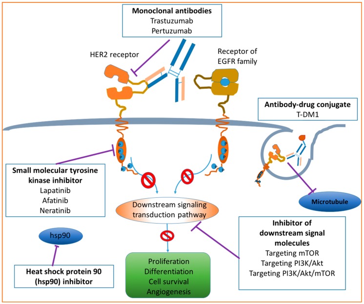
Breast cancer is the second leading cause of cancer death among women. Human epidermal receptor 2 (HER2) positive breast cancer (HER2+ BC) is the most aggressive subtype of breast cancer, with poor prognosis and a high rate of recurrence. About one third of breast cancer is HER2+ BC with significantly high expression level of HER2 protein compared to other subtypes. Therefore, HER2 is an important biomarker and an ideal target for developing therapeutic strategies for the treatment HER2+ BC. In this review, HER2 structure and physiological and pathological roles in HER2+ BC are discussed. Two diagnostic tests, immunohistochemistry (IHC) and fluorescent in situ hybridization (FISH), for evaluating HER2 expression levels are briefly introduced. The current mainstay targeted therapies for HER2+ BC include monoclonal antibodies, small molecule tyrosine kinase inhibitors, antibody-drug conjugates (ADC) and other emerging anti-HER2 agents. In clinical practice, combination therapies are commonly adopted in order to achieve synergistic drug response. This review will help to better understand the molecular mechanism of HER2+ BC and further facilitate the development of more effective therapeutic strategies against HER2+ BC.

Keywords: HER2 positive breast cancer; antibody–drug conjugates; diagnostic tests; molecular mechanism; monoclonal antibodies; small molecular inhibitors; translational therapy.


Conflict of interest statement

The authors declare no conflict of interest.

Figures

Figure 1
Figure 1
Basic structure of epidermal growth factor receptor (EGFR) transmembrane proteins. In the extracellular domain, LD1 and LD2 are two repeated ligand binding domains. CR1 and CR2 are two repeated cysteine rich regions. TM indicates the short transmembrane spanning sequences. In the intracellular domain, TK is a catalytic tyrosine kinase, and CT is the carboxyl-terminal tail. Circled Ps are the phosphorylation sites within the TK and CT regions. This figure is revised based on the review of the oncogene human epidermal growth factor 2 (HER2) contributed by Moasser [17].
Figure 2
Figure 2
Schematic diagram of HER2 signaling pathways. Upon ligand binding, dimerization between receptors of EGFR family and HER2 receptor is induced. The homodimers or heterodimers thereafter stimulate a serial of signaling cascades. Among various signaling pathways, the phosphatidyl inositol 3-kinase (PI3K) and mitogen-activated protein kinase (MAPK) pathways are the two major and most studied pathways which take a pivotal role in tumor proliferation and anti-apoptosis. The whole signal transduction process can be divided into three sections: signal input (ligand-binding and dimerization), signal processing (a series of signaling cascades) and signal output (corresponding cellular processes). The scheme is modified based on two works contributed by Yarden et al. [15] and Tai et al. [16], respectively.
Figure 3
Figure 3
Workflow of HER2 positive breast cancer diagnosis. Tumor samples are initially tested by immunohistochemistry (IHC). Then the samples are divided into three subtypes based on the slide scores of IHC: negative report cases (IHC 0/1+), equivocal cases (IHC 2+) and positive cases (IHC 3+). The equivocal samples will be retested by FISH to verify its HER2 expression more accurately. The figure is a modified version based on Bilous’s work [44].
Figure 4
Figure 4
Molecular approaches for HER2+ BC therapy. Modified from the work contributed by Tsang [13]. Drugs targeting HER2 may include monoclonal antibodies, small molecular tyrosine kinase inhibitors, antibody–drug conjugates, heat shock protein 90 inhibitors and inhibitors of downstream signal molecules. T-DM1: Trastuzumab–emtansine.
Figure 5
Figure 5
Potential action mechanisms of trastuzumab targeting HER2 receptor. (A) Blocking of the dimerization of HER2 and other EGF receptors; (B) Role of antibody-dependent immune-mediated response; (C) Binding of trastuzumab and HER2 may prevent HER2 extracellular domain from cleavage or shedding, which would further inhibit downstream signaling transductions and promote cell apoptosis; and (D) Endocytosis of HER2 receptor conjugated with trastuzumab. Reproduced based on Hudis’s work [59].
Figure 6
Figure 6
Chemical structure of lapatinib, with the chemical formula C29H26ClFN4O4S and a molecular weight of 581.0575 g/mol.
Figure 7
Figure 7
Chemical structure of afatinib, with the chemical formula C24H25ClFN5O3S and a molecular weight of 485.9384 g/mol.
Figure 8
Figure 8
Chemical structure of neratinib, with the chemical formula C30H29ClFN6O3S and a molecular weight of 557.0427 g/mol.
Figure 9
Figure 9
Molecular structure of T-DM1. The monoclonal antibody (trastuzumab) was conjugated with a cytotoxic agent (emtansine, which is also named DM1) through a thioether linker.
Figure 10
Figure 10
Potential action mechanism of T-DM1. (A) Trastuzumab specifically recognizes and binds to extracellular domain of HER2; (B) induced passive endocytosis of the formed complex of trastuzumab and HER2; (C) the complex may undergo a degradation process to separate the DM-1 from the complex; and (D) the liberated DM-1 exerts its cytotoxicity on the microtubule. Modified based on the work contributed by Martinez et al. [94].

References

    1. Centers for Disease Control and Prevention Cancer Among Women. [(accessed on 10 September 2016)]; Available online: http://www.cdc.gov/cancer/dcpc/data/women.htm.
    1. Breastcancer.org U.S. Breast Cancer Statistics. [(accessed on 10 September 2016)]. Available online: http://www.breastcancer.org/symptoms/understand_bc/statistics.
    1. Orphanos G., Kountourakis P. Targeting the HER2 receptor in metastatic breast cancer. Hematol. Oncol. Stem Cell Ther. 2012;5:127–137. doi: 10.5144/1658-3876.2012.127. - DOI - PubMed
    1. Slamon D.J., Clark G.M., Wong S.G., Levin W.J., Ullrich A., McGuire W.L. Human breast cancer: Correlation of relapse and survival with amplification of the HER−2/neu oncogene. Science. 1987;235:177–182. doi: 10.1126/science.3798106. - DOI - PubMed
    1. Slamon D.J. Studies of the HER−2/neu Proto-oncogene in Human Breast Cancer. Cancer Investig. 1990;8:253–254. doi: 10.3109/07357909009017573. - DOI - PubMed

MeSH terms