. 2016 Dec 14;21(12):1718.
doi: 10.3390/molecules21121718.

Scoparone Inhibits LPS-Simulated Inflammatory Response by Suppressing IRF3 and ERK in BV-2 Microglial Cells

Affiliations

Scoparone Inhibits LPS-Simulated Inflammatory Response by Suppressing IRF3 and ERK in BV-2 Microglial Cells

Duk-Yeon Cho et al. Molecules. .

Abstract

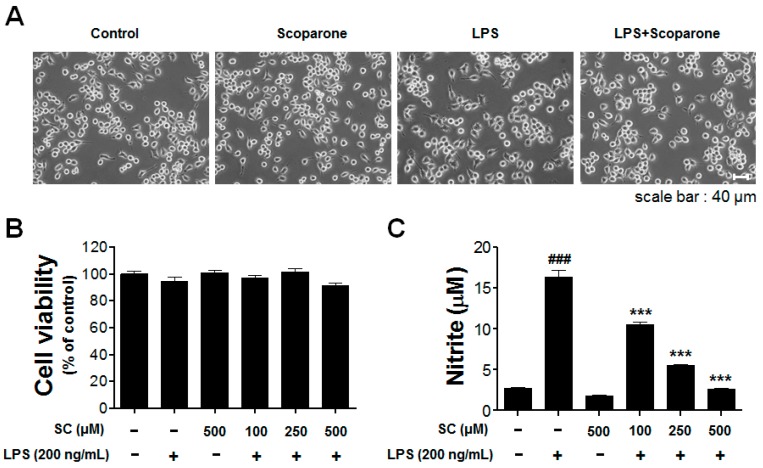
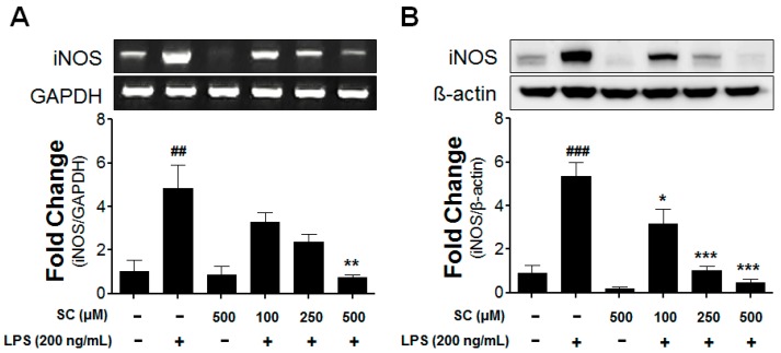
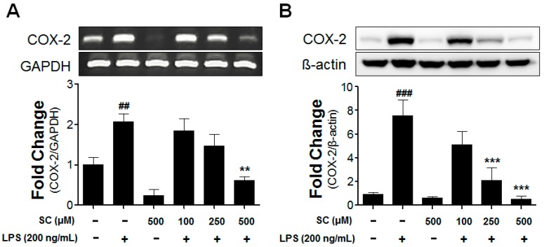
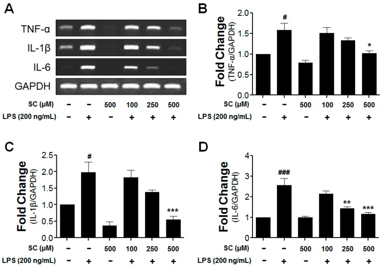
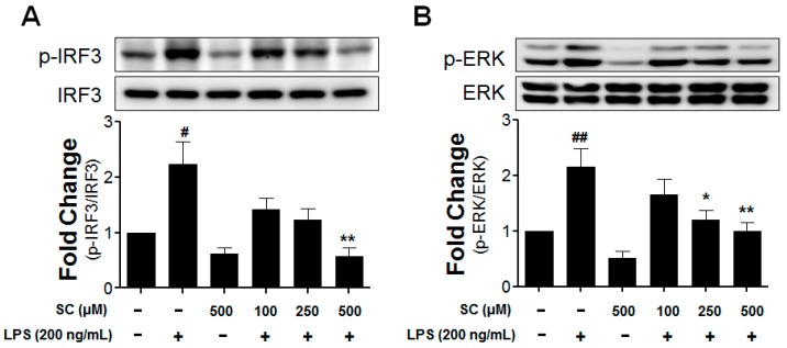
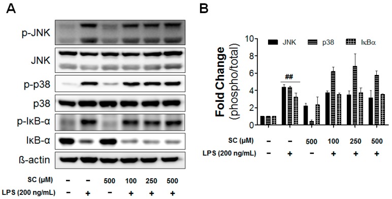
Microglia activation and the release of various inflammatory cytokines are largely related to neurological diseases, including Parkinson's, Alzheimer's, and other brain diseases. The suppression of microglial cells using natural bioactive compounds has become increasingly important for brain therapy owing to the expected beneficial effect of lower toxicity. Scoparone (6,7-dimethoxycoumarin), a major bioactive compound found in various plant parts, including the inner shell of chestnut (Castanea crenata), was evaluated on lipopolysaccharide (LPS)-activated BV-2 microglia cells. The results indicated that scoparone suppresses the LPS-stimulated increase of neuroinflammatory responses and inhibited the pro-inflammatory cytokine production in the BV-2 microglial cells. A mechanistic study showed that scoparone specifically inhibited the LPS-stimulated activation via a major regulation of IRF-3 and a regulation of ERK, whereby the phosphorylation in the BV-2 microglial cells is blocked. These data suggest that scoparone has anti-neuroinflammatory effects in LPS-activated BV-2 microglial cells, and could possibly be used in the development of novel drugs for the prevention and treatment of neuroinflammatory diseases.

Keywords: ERK; IRF-3; microglial cells; scoparone.


Conflict of interest statement

The authors declare no conflicts of interest.

Figures

Figure 1
Figure 1
Effect of scoparone on cell viability and nitric-oxide production in LPS-induced BV-2 microglial cells. The morphological changes are represented in the BV2 microglial cells (A). The viability in the scoparone-treatment cells was evaluated using the MTT assay (B). The BV-2 microglial cells were incubated with 100 μM, 250 μM, and 500 μM of scoparone for 24 h. The results are displayed as a percentage of the control samples. The nitrite in the medium was determined using the Griess assay (C). Data are the mean ± standard error (n = 3) of three independent experiments. ### p < 0.005, compared with the control group; *** p < 0.005 compared with the LPS-treated group.
Figure 2
Figure 2
Effect of scoparone on LPS-induced iNOS-mRNA and iNOS-protein expressions in BV-2 microglial cells. The BV-2 microglial cells were seeded at 2.5 × 10⁵ cells/mL, and were incubated for 6 h and 20 h with various concentrations of scoparone 1 h before the stimulation with LPS. The mRNA was first isolated, and the mRNA expression was then evaluated using the RT-PCR (A). The cell lysates were electrophoresed, and the iNOS expressions were detected using a specific antibody (B). The data are the mean ± standard error (n = 3) of three independent experiments. ## p < 0.01, ### p < 0.005, compared with the control group; * p < 0.05, ** p < 0.01, and *** p < 0.005 compared with the LPS-treated group.
Figure 3
Figure 3
Effect of scoparone on LPS-induced COX-2-mRNA and COX-2-protein expressions in BV-2 microglial cells. The BV-2 microglial cells were seeded at 2.5 × 10⁵ cells/mL, and were incubated for 6 h and 20 h with various concentrations of scoparone 1 h before the stimulation with LPS. The mRNA was first isolated, and the mRNA expression was then evaluated using the RT-PCR (A). The cell lysates were electrophoresed, and the COX-2 expressions were detected using a specific antibody (B). The data are the mean ± standard error (n = 3) of three independent experiments. ## p < 0.01, ### p < 0.005, compared with the control group; ** p < 0.01 and *** p < 0.005 compared with the LPS-treated group.
Figure 4
Figure 4
Effect of scoparone on mRNA expression of LPS-induced cytokines in BV-2 microglial cells. The BV-2 microglial cells were seeded at 2.5 × 10⁵ cells/mL, and were incubated for 6 h with various concentrations of scoparone 1 h before the stimulation with LPS. The mRNA was first isolated, and the mRNA expressions of the tumor-necrosis factor (TNF)-α, interleukin (IL)-1β, and interleukin (IL)-6 were then evaluated using the RT-PCR (A). The representative densitometry analyses of TNF-α (B), IL-1β (C) and IL-6 (D) compared with GAPDH mRNA. The data are the mean ± standard error (n = 3) of three independent experiments. # p < 0.05, ### p < 0.005, compared with the control group; * p < 0.05, ** p < 0.01, and *** p < 0.005 compared with the LPS-treated group.
Figure 5
Figure 5
Effect of scoparone on LPS-induced IRF3 and ERK activations induced by LPS in BV-2 microglial cells. The BV-2 microglial cells were seeded at 2.5 × 10⁵ cells/mL, and were incubated for 1 h with various concentrations of scoparone 1 h before the stimulation with LPS. The cell lysates were electrophoresed, and the phospho-IRF3 (A), phospho-ERK (B), and ERK expressions were detected using a specific antibody. # p < 0.05, ## p < 0.01, compared with the control group; * p < 0.05, ** p < 0.01 compared with the LPS-treated group.
Figure 6
Figure 6
Effect of scoparone on LPS-induced MAPKs and NF-κB activation in BV-2 microglial cells. The BV-2 microglial cells were seeded at 2.5 × 10⁵ cells/mL, and were incubated for 1 h with various concentrations of scoparone 1 h before the stimulation with LPS. The cell lysates were electrophoresed, and the phospho-JNK, JNK, phospho-p38, p38 MAPKs, phospho-IκB-α, and IκB-α expressions were detected using a specific antibody (A). The quantification data are shown in the right panel (B). Data are the mean ± standard error (n = 3) of three independent experiments. ## p < 0.01, compared with the control group.

References

    1. Graeber M.B., Li W., Rodriguez M.L. Role of microglia in CNS inflammation. FEBS Lett. 2011;585:3798–3805. doi: 10.1016/j.febslet.2011.08.033. - DOI - PubMed
    1. Von Bernhardi R., Tichauer J.E., Eugenin J. Aging-dependent changes of microglial cells and their relevance for neurodegenerative disorders. J. Neurochem. 2010;112:1099–1114. doi: 10.1111/j.1471-4159.2009.06537.x. - DOI - PubMed
    1. Kaur G., Han S.J., Yang I., Crane C. Microglia and central nervous system immunity. Neurosurg. Clin. N. Am. 2010;21:43–51. doi: 10.1016/j.nec.2009.08.009. - DOI - PubMed
    1. Kim B.W., Koppula S., Kumar H., Park J.Y., Kim I.W., More S.V., Kim I.S., Han S.D., Kim S.K., Yoon S.H., et al. Alpha-Asarone attenuates microglia-mediated neuroinflammation by inhibiting NF kappa B activation and mitigates MPTP-induced behavioral deficits in a mouse model of Parkinson’s disease. Neuropharmacology. 2015;97:46–57. doi: 10.1016/j.neuropharm.2015.04.037. - DOI - PubMed
    1. Lim H.W., Park J.I., More S.V., Park J.Y., Kim B.W., Jeon S.B., Yun Y.S., Park E.J., Yoon S.H., Choi D.K. Anti-neuroinflammatory effects of DPTP, a novel synthetic clovamide derivative in in vitro and in vivo model of neuroinflammation. Brain Res. Bull. 2015;112:25–34. doi: 10.1016/j.brainresbull.2015.01.004. - DOI - PubMed

MeSH terms

LinkOut - more resources