. 2016 Dec 15;21(12):1725.
doi: 10.3390/molecules21121725.

Half-Sandwich Ru(II) Halogenido, Valproato and 4-Phenylbutyrato Complexes Containing 2,2'-Dipyridylamine: Synthesis, Characterization, Solution Chemistry and In Vitro Cytotoxicity

Affiliations

Half-Sandwich Ru(II) Halogenido, Valproato and 4-Phenylbutyrato Complexes Containing 2,2'-Dipyridylamine: Synthesis, Characterization, Solution Chemistry and In Vitro Cytotoxicity

Pavel Štarha et al. Molecules. .

Abstract

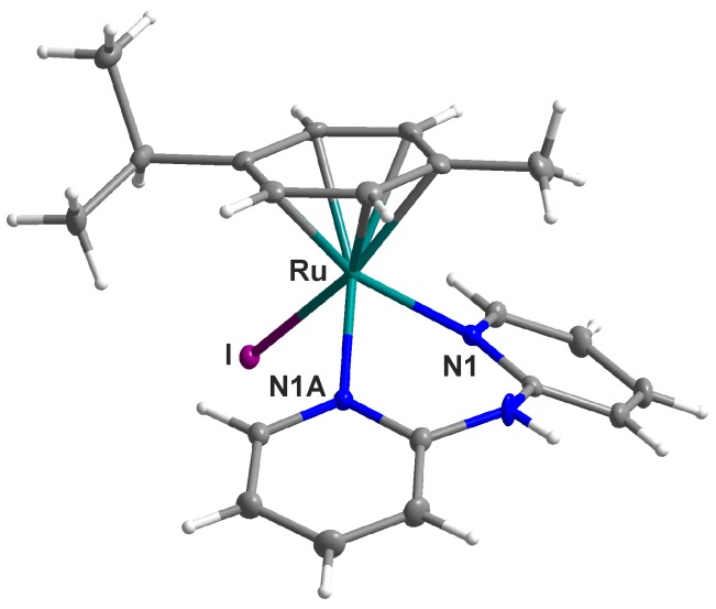
Halogenido and carboxylato Ru(II) half-sandwich complexes of the general composition [Ru(η⁶-p-cym)(dpa)X]PF₆ (1-5) were prepared and thoroughly characterized with various techniques (e.g., mass spectrometry, NMR spectroscopy and X-ray analysis); dpa = 2,2'-dipyridylamine; p-cym = p-cymene; X = Cl- (for 1), Br- (for 2), I- (for 3), valproate(1-) (for 4) or 4-phenylbutyrate(1-) (for 5). A single-crystal X-ray analysis showed a pseudo-octahedral piano-stool geometry of [Ru(η⁶-p-cym)(dpa)I]PF₆ (3), with a η⁶-coordinated p-cymene, bidentate N-donor dpa ligand and iodido ligand coordinated to the Ru(II) atom. The results of the ¹H-NMR solution behaviour studies proved that the complexes 1-5 hydrolyse were in the mixture of solvents used (10% MeOD-d₄/90% D₂O). Complexes 1-5 were in vitro inactive against the A2780 human ovarian carcinoma cell line, up to the highest tested concentration (IC50 > 100 μM).

Keywords: 2,2′-dipyridylamine; X-ray structure; half-sandwich; in vitro cytotoxicity; ruthenium; solution behaviour.


Conflict of interest statement

The authors declare no conflict of interest.

Figures

Figure 1
Figure 1
General structural formula of the studied complexes 15 given with the atom numbering scheme for the p-cym and dpa ligands; p-cym = p-cymene; dpa = 2,2′-dipyridylamine; X = Cl (1), Br (2), I (3), valproate(1−) (4) or 4-phenylbutyrate(1−) (5).
Figure 2
Figure 2
ESI+ mass spectra of the carboxylato complexes [Ru(η6-p-cym)(dpa)(VP)]PF6 (4; top) and [Ru(η6-p-cym)(dpa)(PB)]PF6 (5; bottom), given with a comparison of the experimental and simulated isotopic distributions of the [Ru(p-cym)(dpa)(VP)]+ and [Ru(p-cym)(dpa)(PB)]+ species (insets).
Figure 3
Figure 3
1H-NMR spectra of [Ru(η6-p-cym)(dpa)Cl]PF6 (1; top) and [Ru(η6-p-cym)(dpa)VP]PF6 (4; bottom), given together with the general assignment of the observed signals, as follows; green dots for the dpa signals, blue dots for the p-cym signals and red dots for the VP signals.
Figure 4
Figure 4
Molecular structure of [Ru(η6-p-cym)(dpa)I]PF6 (3). Non-hydrogen atoms are drawn as thermal ellipsoids at the 50% probability level. The PF6 counterion has been omitted for clarity.
Figure 5
Figure 5
Parts of the 1H-NMR spectra of complex 1 and its mixture with 2 molar equivalents of the reduced glutathione (GSH), both after 48 h of standing at ambient temperature. 1H-NMR spectrum of free GSH is given for comparative purposes.
Figure 6
Figure 6
A schema of the hydrolysis of complex [Ru(η6-p-cym)(dpa)(VP)]PF6 (4) given together with the tentative compositions of the hydrolysates, i.e., the [Ru(η6-p-cym)(dpa)(H2O)]2+ and [Ru(η6-p-cym)(dpa)(OH)]+ species (top). 1H-NMR spectra of the representative signal of the terminal C25–H methyl group of released valproate(1–) anion (ca. 0.78 ppm; red) and the valproato ligand (ca. 0.62 ppm; green), as observed for complex 4, without or with the addition of the reduced glutathione (GSH) at different time points. 1H-NMR spectrum of free sodium valproate (NaVP) is given for comparative purposes (bottom).
Figure 7
Figure 7
Progress of hydrolysis of the carboxylato complexes 4 (blue) and 5 (dark yellow), with (circles) or without (squares) the addition of 2 molar equivalents of the reduced glutathione (GSH). Complexes were dissolved in 10% MeOD-d4/90% D2O and the 1H-NMR spectra were recorded on the fresh solutions (0 h) and after 0.5, 1, 2, 4, 6, 24 and 48 h of standing at ambient temperature.
Figure 8
Figure 8
Structural formula of the complex cation in [Ru(η6-p-cym)(dpa)Cl]PF6 (1).
Figure 9
Figure 9
Structural formula of the complex cation in [Ru(η6-p-cym)(dpa)Br]PF6 (2).
Figure 10
Figure 10
Structural formula of the complex cation in [Ru(η6-p-cym)(dpa)I]PF6 (3).
Figure 11
Figure 11
Structural formula of the complex cation in [Ru(η6-p-cym)(dpa)(VP)]PF6 (4); VP = valproato(1–).
Figure 12
Figure 12
Structural formula of the complex cation in [Ru(η6-p-cym)(dpa)(PB)]PF6 (5); PB = 4-phenylbutyrato(1–).

References

    1. Murray B.S., Babak M.V., Hartinger C.G., Dyson P.J. The development of RAPTA compounds for the treatment of tumors. Coord. Chem. Rev. 2016;306:86–114. doi: 10.1016/j.ccr.2015.06.014. - DOI
    1. Süss-Fink G. Arene ruthenium complexes as anticancer agents. Dalton Trans. 2010;39:1673–1688. doi: 10.1039/B916860P. - DOI - PubMed
    1. Romero-Canelón I., Salassa L., Sadler P.J. The contrasting activity of iodido versus chlorido ruthenium and osmium arene azo- and imino-pyridine anticancer complexes: Control of cell selectivity, cross-resistance, p53 dependence, and apoptosis pathway. J. Med. Chem. 2013;56:1291–1300. doi: 10.1021/jm3017442. - DOI - PubMed
    1. Chiu C.T., Wang Z., Hunsberger J.G., Chuang D.M. Therapeutic potential of mood stabilizers lithium and valproic acid: Beyond bipolar disorder. Pharmacol. Rev. 2013;65:105–142. doi: 10.1124/pr.111.005512. - DOI - PMC - PubMed
    1. Iannitti T., Palmieri B. Clinical and experimental applications of sodium phenylbutyrate. Drugs R&D. 2011;11:227–249. - PMC - PubMed

LinkOut - more resources