. 2017 Jan 20;12(1):282-290.
doi: 10.1021/acschembio.6b00791. Epub 2016 Dec 16.

Internal Structure and Preferential Protein Binding of Colloidal Aggregates

Affiliations

Internal Structure and Preferential Protein Binding of Colloidal Aggregates

Da Duan et al. ACS Chem Biol. .

Abstract

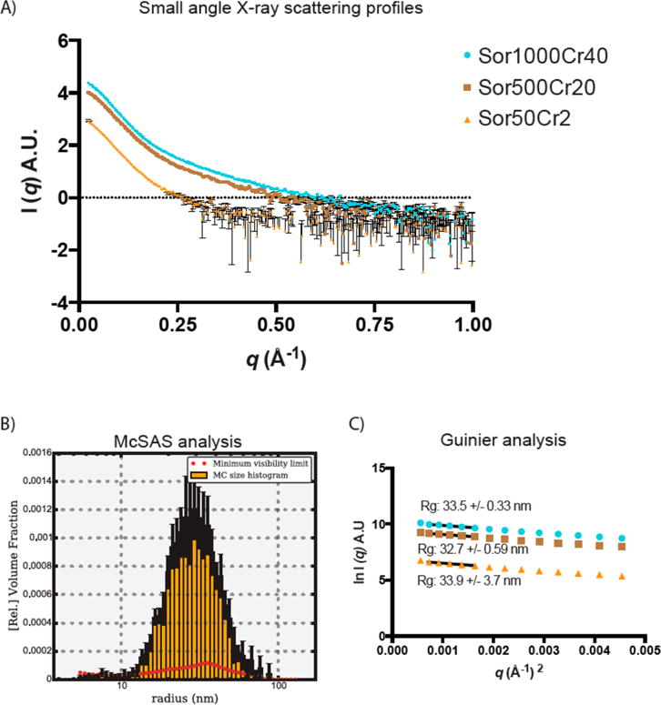
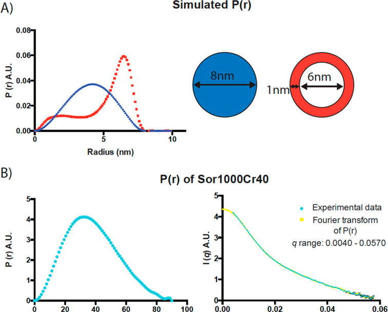
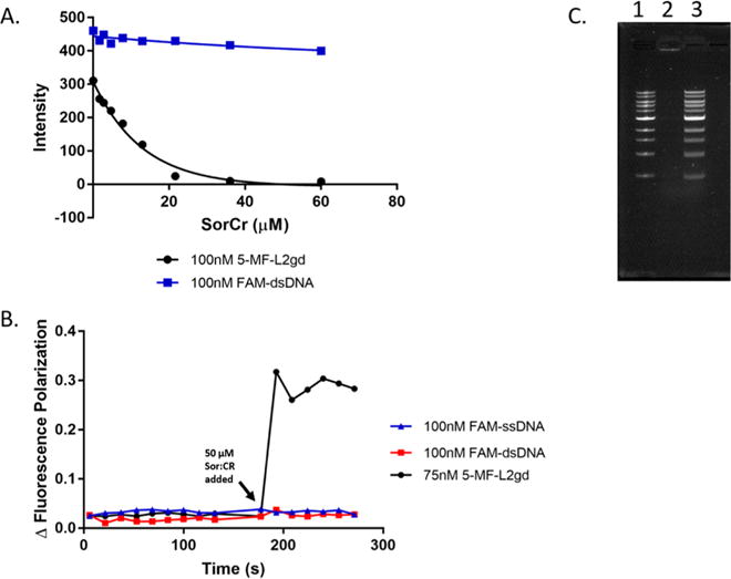
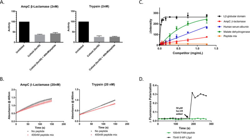
Colloidal aggregates of small molecules are the most common artifact in early drug discovery, sequestering and inhibiting target proteins without specificity. Understanding their structure and mechanism has been crucial to developing tools to control for, and occasionally even exploit, these particles. Unfortunately, their polydispersity and transient stability have prevented exploration of certain elementary properties, such as how they pack. Dye-stabilized colloidal aggregates exhibit enhanced homogeneity and stability when compared to conventional colloidal aggregates, enabling investigation of some of these properties. By small-angle X-ray scattering and multiangle light scattering, pair distance distribution functions suggest that the dye-stabilized colloids are filled, not hollow, spheres. Stability of the coformulated colloids enabled investigation of their preference for binding DNA, peptides, or folded proteins, and their ability to purify one from the other. The coformulated colloids showed little ability to bind DNA. Correspondingly, the colloids preferentially sequestered protein from even a 1600-fold excess of peptides that are themselves the result of a digest of the same protein. This may reflect the avidity advantage that a protein has in a surface-to-surface interaction with the colloids. For the first time, colloids could be shown to have preferences of up to 90-fold for particular proteins over others. Loaded onto the colloids, bound enzyme could be spun down, resuspended, and released back into buffer, regaining most of its activity. Implications of these observations for colloid mechanisms and utility will be considered.


Conflict of interest statement

Notes

The authors declare no competing financial interest.

Figures

Figure 1
Figure 1
Small-angle X-ray scattering from the coformulated colloids supporting a more homogeneous population of particles. (A) SAXS scattering profiles of Sor/CR colloids at three different concentrations within q range of 0.028–1 Å−1. SAXS scattering profiles of Sor/CR (25:1, 1000 μM sorafenib), Sor/CR (25:1, 500 μM sorafenib), and Sor/CR (25:1, 50 μM sorafenib) are in cyan, brown, and orange, respectively. (B) McSAS anaylsis of SAXS scattering profile of Sor/CR (25:1, 1000 μM sorafenib) colloids using only the first 170 data points. (C) Guinier analysis using only data points 2–6 of all three scattering profiles to obtain radius of gyration. All spectra taken in 50 mM KPi, pH 7.0.
Figure 2
Figure 2
The SAXS spectra, supporting a filled core structure for the colloidal particles. (A) Simulated P(r) functions for two particles of the same diameter but different cores-shell structures. (B) Left panel is P(r) function obtained from SAXS scattering profile of Sor/CR (25:1; 1000 μM sorafenib) colloids in the q range of 0.0040–0.0570 Å−1. Right panel shows a good fit between Fourier transformed P(r) in the left panel and the raw SAXS scattering data, indicating that the P(r) is an accurate representation of the scattering profile.
Figure 3
Figure 3
Colloids preferentially bind protein versus DNA (A) The fluorescence intensity of fluorescein-labeled L2gd protein (5-MF-L2gd, black curve), but not fluorescein labeled dsDNA (FAM-dsDNA, blue curve), is quenched in the presence of increasing amounts of Sor/CR colloidal aggregates. (B) Whereas the addition of 50 μM Sor/CR colloids significantly increases the polarization of 5-MF-L2gd protein, the colloids had little measurable effect on the polarization of dsDNA or ssDNA also labeled with fluorescein. (C) Incubation with 1 Kb DNA ladder (lane 1, precolloids) and subsequent spin-down and resuspension of the colloidal pellet yields no DNA (lane 2). Instead, DNA is detected in the supernatant (lane 3).
Figure 4
Figure 4
Colloids preferentially protein versus peptides. (A) The amounts of inhibited activity of 2 nM of AmpC β-lactamase or 2 nM trypsin by Sor/CR (25:1, 50 μM sorafenib) colloids in the presence and absence of 400 nM peptide mixture do not exhibit major differences. (B) Spin-down experiment where 20 nM of AmpC β-lactamase or trypsin was incubated with Sor/CR (25:1, 500 μM sorafenib) for 5 min in the presence and absence of 3.2 μM of the eight peptide mixture (400 nM in each of the eight peptides). The solution is centrifuged, and the resulting pellet is resuspended in buffer. Colloid disruption by 0.01% Triton X-100 releases the enzymes back into solution, and only slight differences are observed between the activity of samples with and without the peptide mixture. (C) The competitive displacement of labeled 5-MF-L2gd on the colloid by other proteins increases its fluorescence. Shown is competition with unlabeled L2gd itself, human serum albumin, malate dehydrogenase, and AmpC β-lactamase. The large range in apparent IC50 values supports differential binding by different proteins. Meanwhile, the eight peptide mixture has little measurable ability to compete with protein for colloid binding. (D) Consistent with this observation, the addition of 50 μM Sor/CR colloids substantially increases the polarization of 5-MF-L2gd protein but had little measurable effect on the polarization of the labeled HTFPAVL peptide, suggesting that the latter does not bind to the colloid.

References

    1. McGovern SL, Caselli E, Grigorieff N, Shoichet BK. A common mechanism underlying promiscuous inhibitors from virtual and high-throughput screening. J Med Chem. 2002;45:1712–1722. - PubMed
    1. Coan KE, Shoichet BK. Stoichiometry and physical chemistry of promiscuous aggregate-based inhibitors. J Am Chem Soc. 2008;130:9606–9612. - PMC - PubMed
    1. Wolan DW, Zorn JA, Gray DC, Wells JA. Small-molecule activators of a proenzyme. Science. 2009;326:853–858. - PMC - PubMed
    1. McGovern SL, Helfand B, Feng B, Shoichet BK. A Specific Mechanism for Non-Specific Inhibition. J Med Chem. 2003;46:4265–4272. - PubMed
    1. Ryan AJ, Gray NM, Lowe PN, Chung C. Effect of Detergent on “Promiscuous” Inhibitors. J Med Chem. 2003;46:3448–3451. - PubMed

MeSH terms