. 2016 Dec 16;12(12):e1006475.
doi: 10.1371/journal.pgen.1006475. eCollection 2016 Dec.

The C. elegans Discoidin Domain Receptor DDR-2 Modulates the Met-like RTK-JNK Signaling Pathway in Axon Regeneration

Affiliations

The C. elegans Discoidin Domain Receptor DDR-2 Modulates the Met-like RTK-JNK Signaling Pathway in Axon Regeneration

Naoki Hisamoto et al. PLoS Genet. .

Abstract

The ability of specific neurons to regenerate their axons after injury is governed by cell-intrinsic regeneration pathways. However, the signaling pathways that orchestrate axon regeneration are not well understood. In Caenorhabditis elegans, initiation of axon regeneration is positively regulated by SVH-2 Met-like growth factor receptor tyrosine kinase (RTK) signaling through the JNK MAPK pathway. Here we show that SVH-4/DDR-2, an RTK containing a discoidin domain that is activated by collagen, and EMB-9 collagen type IV regulate the regeneration of neurons following axon injury. The scaffold protein SHC-1 interacts with both DDR-2 and SVH-2. Furthermore, we demonstrate that overexpression of svh-2 and shc-1 suppresses the delay in axon regeneration observed in ddr-2 mutants, suggesting that DDR-2 functions upstream of SVH-2 and SHC-1. These results suggest that DDR-2 modulates the SVH-2-JNK pathway via SHC-1. We thus identify two different RTK signaling networks that play coordinated roles in the regulation of axonal regeneration.


Conflict of interest statement

The authors have declared that no competing interests exist.

Figures

Fig 1
Fig 1. DDR-2 is required for efficient axon regeneration in C. elegans.
(A) SVH-2–JNK MAPK pathway required for axon regeneration in C. elegans. The growth factor SVH-1 and its receptor tyrosine kinase SVH-2 promote axon regeneration through tyrosine phosphorylation of MLK-1 in the KGB-1 JNK pathway. (B) Structures of DDR-1 and DDR-2. Schematic diagrams of DDR-1, DDR-2 and their mammalian counterpart DDR2 are shown. Domains are shown as follows: a signal sequence (SS), a discoidin domain (DS), a DS-like domain, a transmembrane domain (TM), and a tyrosine kinase domain (Kinase). The bold lines underneath indicate the extent of the deleted region in each deletion mutant. The ddr-2(tm797) mutation causes a premature translation stop (indicated by asterisk) in the extracellular domain. (C) Representative D-type motor neurons in wild-type and ddr-2 mutant animals 24 hr after laser surgery. In wild-type animals, a severed axon has regenerated a growth cone (arrow). In ddr-2 mutants, proximal ends of axons failed to regenerate (arrowheads). Scale bar = 10 μm. (D,F) Percentages of axons that initiated regeneration 24 hr after laser surgery. Error bars indicate 95% CI. *P<0.05, ***P<0.001. NS, not significant. (E) Percentages of axons that initiated regeneration 24 or 72 hr after laser surgery. Error bars indicate 95% CI. **P<0.01, ***P<0.001. NS, not significant.
Fig 2
Fig 2. Tyrosine kinase activity of DDR-2.
(A) Schematic diagrams of DDR-2, DDR-2C and Tpr-DDR-2C. LZ, leucine zipper. (B,C) Auto-tyrosine-phosphorylation of DDR-2. COS-7 cells were transfected with control vector or the plasmids encoding FLAG-DDR-2C, FLAG-Tpr-DDR-2C (WT) and FLAG-Tpr-DDR-2C (K554E) (KN), as indicated. Cell lysates were immunoprecipitated with anti-FLAG antibody (IP: FLAG) and immunoblotted with anti-phospho-tyrosine (pY) and anti-FLAG antibodies.
Fig 3
Fig 3. Localization of DDR-2.
(A,B) Localization of DDR-2::GFP in D-type motor neurons after axon injury. Fluorescent images of severed axons in wild-type animals carrying Punc-25::ddr-2::gfp and Punc-47::mcherry are shown. Each image was taken at the indicated times after laser surgery. Arrowheads indicate the ends of proximal axons (A). The image was taken at 4 hr after axon injury. Arrows indicate the tip of the proximal injured axon (B). Scale bars = 2 μm. (C) Quantification of the relative fluorescence levels of DDR-2::GFP on the tips of the severed axons. The relative fluorescent intensities of severed axons in animals carrying Punc-25::ddr-2::gfp and Punc-47::mcherry::caax were compared. “N” = number of axons. Error bars indicate 95% CI. *P<0.05, **P<0.01.
Fig 4
Fig 4. The relationship between DDR-2 and SVH-2 in axon regeneration.
(A,C) Percentages of axons that initiated regeneration 24 hr after laser surgery. Error bars indicate 95% CI. *P<0.05, **P<0.01, ***P<0.001. NS, not significant. (B) Tyrosine phosphorylation of MLK-1. COS-7 cells were transfected with plasmids encoding FLAG-Tpr-SVH-2C, FLAG-Tpr-DDR-2C and HA-MLK-1, as indicated. Complexes immunoprecipitated with anti-HA or anti-FLAG antibody were analyzed by immunoblotting with anti-phospho-tyrosine (pY), anti-HA and anti-FLAG antibodies.
Fig 5
Fig 5. Interactions of SHC-1 with SVH-2 and DDR-2.
(A) The relationship among EMB-9–DDR-2, SHC-1 and SVH-1–SVH-2 in the JNK signaling pathway. (B) Structure of SHC-1. Dark and hatched boxes represent the PTB and SH2 domains, respectively. Essential Arg residues required for binding to phospho-tyrosine in PTB and SH2 domains are indicated by asterisks. (C,D) Interactions of SHC-1 with SVH-2 and DDR-2. COS-7 cells were transfected with plasmids encoding FLAG-Tpr-SVH-2C (WT), FLAG-Tpr-SVH-2C(K767R) (KN), FLAG-Tpr-DDR-2C (WT), FLAG-Tpr-DDR-2C (K554E) (KN), T7-SHC-1 (WT), T7-SHC-1 (R136K), T7-SHC-1 (R234K) and T7-SHC-1 (R136K; R234K), as indicated. Whole-cell extracts and immunoprecipitated complexes obtained with anti-FLAG antibody (IP: FLAG) were analyzed by immunoblotting. (E) Percentages of axons that initiated regeneration 24 hr after laser surgery. Error bars indicate 95% CI. **P<0.01, ***P<0.001. NS, not significant.
Fig 6
Fig 6. DDR-2 modulates SVH-1–SVH-2 signaling to regulate axon regeneration after neuron injury.
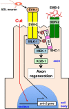
HGF/plasminogen-like protein SVH-1 is constitutively expressed in, and secreted from ADL sensory neurons in the head. Expression of SVH-2 is induced by axon injury. Following axon injury, DDR-2 accumulates at the severed end and is activated by EMB-9. DDR-2 facilitates the efficient activation of SVH-2 by SVH-1. Activated SVH-2 phosphorylates MLK-1 MAPKKK at a tyrosine residue, creating a docking site for SHC-1. SHC-1 constitutively forms a complex with MEK-1 MAPKK, and thereby functions to connect MLK-1 and MEK-1. SHC-1 participates in restricting the SVH-1–SVH-2 signal to the MLK-1–MEK-1 pathway.

Similar articles

Cited by

References

    1. Chen ZL, Yu WM, Strickland S (2007) Peripheral regeneration. Annu Rev Neurosci 30: 209–233. 10.1146/annurev.neuro.30.051606.094337 - DOI - PubMed
    1. Yanik MF, Cinar H, Cinar HN, Chisholm AD, Jin Y, et al. (2004) Neurosurgery: functional regeneration after laser axotomy. Nature 432: 822 10.1038/432822a - DOI - PubMed
    1. O’Brien GS, Sagasti A (2009) Fragile axons forge the path to gene discovery: a MAP kinase pathway regulates axon regeneration. Sci Signal 2: e30. - PubMed
    1. Nix P, Hisamoto N, Matsumoto K, Bastiani M (2011) Axon regeneration requires coordinate activation of p38 and JNK MAPK pathways. Proc Natl Acad Sci USA 108: 10738–10743. 10.1073/pnas.1104830108 - DOI - PMC - PubMed
    1. Camps M, Nichols A, Arkinstall S (2000) Dual specificity phosphatases: a gene family for control of MAP kinase function. FASEB J 14: 6–16. - PubMed

MeSH terms

Substances