

Co-occurrence of histone H3 K27M and BRAF V600E mutations in paediatric midline grade I ganglioglioma
- PMID: 27984673
- PMCID: PMC8028391
- DOI: 10.1111/bpa.12473
Co-occurrence of histone H3 K27M and BRAF V600E mutations in paediatric midline grade I ganglioglioma
Abstract
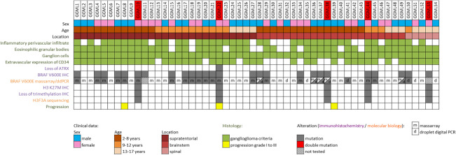
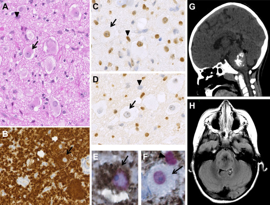
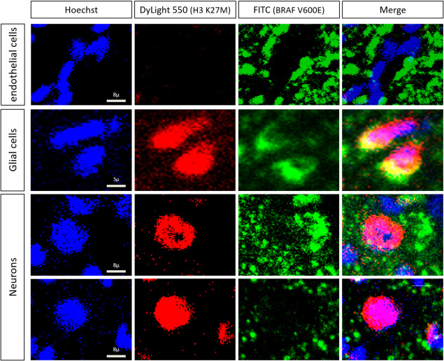
Ganglioglioma (GG) is a grade I tumor characterized by alterations in the MAPK pathway, including BRAF V600E mutation. Recently, diffuse midline glioma with an H3 K27M mutation was added to the WHO 2016 classification as a new grade IV entity. As co-occurrence of H3 K27M and BRAF V600E mutations has been reported in midline tumors and anaplastic GG, we searched for BRAF V600E and H3 K27M mutations in a series of 54 paediatric midline grade I GG (midline GG) to determine the frequency of double mutations and its relevance for prognosis. Twenty-seven patients (50%) possessed the BRAF V600E mutation. The frequency of the co-occurrence of H3F3A/BRAF mutations at diagnosis was 9.3%. No H3 K27M mutation was detected in the absence of the BRAF V600E mutation. Double-immunostaining revealed that BRAF V600E and H3 K27M mutant proteins were present in both the glial and neuronal components. Immunopositivity for the BRAF V600E mutant protein correlated with BRAF mutation status as detected by massARRAY or digital droplet PCR. The median follow-up of patients with double mutation was 4 years. One patient died of progressive disease 8 years after diagnosis, whereas the four other patients were all alive with stable disease at the last clinical follow-up (at 9 months, 1 year and 7 years) without adjuvant therapy. We demonstrate in this first series of midline GGs that the H3 K27M mutation can occur in association with the BRAF V600E mutation in grade I glioneuronal tumors. Despite the presence of H3 K27M mutations, these cases should not be graded and treated as grade IV tumors because they have a better spontaneous outcome than classic diffuse midline H3 K27M-mutant glioma. These data suggest that H3 K27M cannot be considered a specific hallmark of grade IV diffuse gliomas and highlight the importance of integrated histomolecular diagnosis in paediatric brain tumors.
Keywords: BRAF V600E; H3 K27M; ganglioglioma; midline.
© 2016 International Society of Neuropathology.
Conflict of interest statement
The authors declare that they have no conflict of interest except Pascale Varlet (Hoffmann La roche, Novartis and Boehringer‐Ingelheim) and Jacques Grill (Novartis, Roche, Bristol‐Myers Squibb).
Figures
References
-
- Blümcke I, Wiestler OD (2002) Gangliogliomas: an intriguing tumor entity associated with focal epilepsies. J Neuropathol Exp Neurol 61:575–584. - PubMed
-
- Compton JJ, Laack NNI, Eckel LJ, Schomas DA, Giannini C, Meyer FB (2012) Long‐term outcomes for low‐grade intracranial ganglioglioma: 30‐year experience from the Mayo Clinic. J Neurosurg 117:825–830. - PubMed
-
- Dahiya S, Haydon DH, Alvarado D, Gurnett CA, Gutmann DH, Leonard JR (2013) BRAF(V600E) mutation is a negative prognosticator in pediatric ganglioglioma. Acta Neuropathol (Berl) 125:901–910. - PubMed
MeSH terms
Substances
LinkOut - more resources
Full Text Sources
Other Literature Sources
Medical
Research Materials
