Comment
. 2016 Dec 15;167(7):1829-1838.e9.
doi: 10.1016/j.cell.2016.11.017. Epub 2016 Dec 8.

Naturally Occurring Off-Switches for CRISPR-Cas9

Affiliations
Comment

Naturally Occurring Off-Switches for CRISPR-Cas9

April Pawluk et al. Cell. .

Abstract

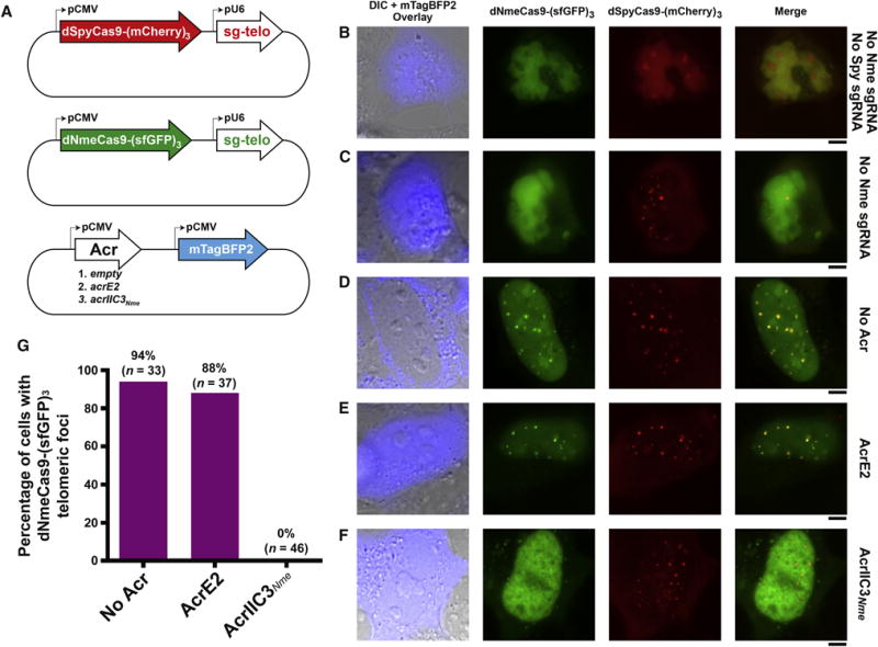
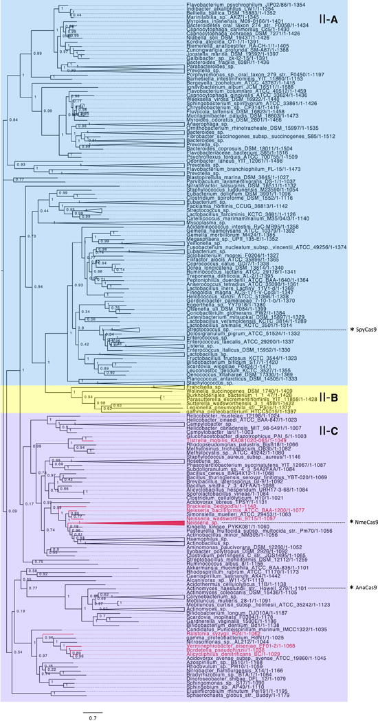
CRISPR-Cas9 technology would be enhanced by the ability to inhibit Cas9 function spatially, temporally, or conditionally. Previously, we discovered small proteins encoded by bacteriophages that inhibit the CRISPR-Cas systems of their host bacteria. These "anti-CRISPRs" were specific to type I CRISPR-Cas systems that do not employ the Cas9 protein. We posited that nature would also yield Cas9 inhibitors in response to the evolutionary arms race between bacteriophages and their hosts. Here, we report the discovery of three distinct families of anti-CRISPRs that specifically inhibit the CRISPR-Cas9 system of Neisseria meningitidis. We show that these proteins bind directly to N. meningitidis Cas9 (NmeCas9) and can be used as potent inhibitors of genome editing by this system in human cells. These anti-CRISPR proteins now enable "off-switches" for CRISPR-Cas9 activity and provide a genetically encodable means to inhibit CRISPR-Cas9 genome editing in eukaryotes. VIDEO ABSTRACT.

Keywords: CRISPR-Cas; Cas9; Neisseria meningitidis; anti-CRISPR; genome editing; phage.


Figures

Figure 1
Figure 1. Identification and Validation of Type II-C Anti-CRISPRs
(A) Schematic representation of candidate type II-C acr and aca genes within putative MGEs in the genomes of strains of Brackiella oedipodis and Neisseria meningitidis. Homologous genes are color-matched, with percent amino acid identities indicated. Gene arrows are not drawn to scale. Any known, relevant gene product functions are annotated as follows: Rep, plasmid replication protein; Reg, transcriptional regulator; Tra, conjugal transfer protein; Rec, recombinase; Tail, phage tail structural protein; Lysis, phage lysis cassette. Genes colored in grey have MGE-related functions and/or show clear evidence of horizontal transfer. (B) Schematic representation of genotypes in N. meningitidis strains used to test candidate anti-CRISPR function. Diamonds, CRISPR repeats; numbered rectangles, CRISPR spacers; arrows, CRISPR transcription. ermC, integrated erythromycin resistance cassette; acrX, integrated candidate anti-CRISPR cassette. Individual genetic elements are not to scale. (C) Candidate type II-C anti-CRISPRs inhibit CRISPR interference in N. meningitidis. Results of the transformation assay in N. meningitidis strain 8013, and isogenic derivatives with each indicated acr gene integrated at the nics locus (see B), are plotted. The CRISPR-targeted protospacer plasmid (yellow) cannot transform wild-type and empty vector-containing cells due to an active CRISPR-Cas system, resulting in zero transformants. BDL = below detection limit of this assay. Plasmid DNA that lacks a target protospacer sequence can transform all strains equally well (navy). Experiments were repeated three times and error bars represent the standard error of the mean (s.e.m) between three replicates. Cells were also plated on non-selective media and the total number of cfu/mL present was equivalent in each sample (data not shown).
Figure 2
Figure 2. Anti-CRISPRs Bind Directly to NmeCas9:sgRNA
(A) Purified, untagged anti-CRISPR proteins were mixed with purified, 6xHis tagged NmeCas9:sgRNA in vitro. The input and elution fractions (before and after nickel affinity purification) are shown on the right and left sides of the Coomassie-stained SDS-PAGE gel, respectively. Mobilities of marker proteins (in kDa) are denoted on the left. AcrE2 is an inhibitor of the type I–E CRISPR-Cas system, and is included in this assay as a negative control. The gel image was cropped to conserve space and to remove irrelevant bands resulting from Cas9 degradation. The image is representative of at least three replicates. Uncropped gel images are presented in Figure S2. (B) Binding assays were carried out between the same anti-CRISPRs tested in (A) and Cas9 from Actinomyces naeslundii (AnaCas9). AnaCas9 is a distantly related type II-C Cas9 protein (~20% amino acid sequence identity with NmeCas9). The image is representative of at least three replicates.
Figure 3
Figure 3. Type II-C Anti-CRISPRs Specifically Block DNA Cleavage by NmeCas9 In Vitro
Linearized plasmid DNA bearing a protospacer adjacent to a PAM sequence was subjected to in vitro digestion by purified, recombinant, sgRNA-programmed NmeCas9 (upper panel) or SpyCas9 (lower panel). Where indicated at the top of each lane, Cas9 was pre-incubated with purified anti-CRISPR proteins as indicated with AcrE2 as a negative control. Molar equivalents of anti-CRISPR protein (relative to Cas9) are shown at the top of each lane, and mobilities of input and cleaved DNAs are denoted on the right. The NmeCas9 cleavage assays shown are representative of three independent replicates.
Figure 4
Figure 4. Type II-C Anti-CRISPRs Specifically Block Genome Editing by NmeCas9 in Human Cells
(A) Schematic representation of R-loop structures at a dual target site (DTS3) in the human genome that can be cleaved and edited by either SpyCas9 (top) or NmeCas9 (bottom). Guide sequences (purple), PAMs (boxed), and Cas9 cleavage sites (red line) are indicated. (B) T7E1 assays of NmeCas9 or SpyCas9 editing efficiencies at DTS3 upon transient transfection of human HEK293T cells. Constructs encoding anti-CRISPR proteins were co-transfected as indicated at the top of each lane. Mobilities of T7E1-digested (edited) and -undigested (unedited) bands are indicated to the right, and editing efficiencies (“% lesion”) are given at the bottom of each lane. These images are representative of at least seven replicates.
Figure 5
Figure 5. AcrIIC3Nme Prevents DNA Binding by NmeCas9 in Human Cells
(A) Schematic representation of plasmids used for expression of dNmeCas9-(sfGFP)3, dSpyCas9-(mCherry)3, their respective telomeric sgRNAs, and anti-CRISPR protein. The plasmid encoding the anti-CRISPR protein is also marked with the blue fluorescent protein mTagBFP2. (B-F) Fluorescence images of U2OS cells transiently transfected with plasmids depicted in (A). The specific version of each plasmid set (with or without sgRNAs, with or without anti-CRISPRs) is given to the right of each row. First column: differential interference contrast (DIC) and mTagBFP2 imaging, merged. Second column: dNmeCas9-(sfGFP)3. Third column: dSpyCas9-(mCherry)3. Fourth column: dNmeCas9-(sfGFP)3 and dSpyCas9-(mCherry)3, merged. Scale bars, 5 μm. (G) Quantitation of dNmeCas9-(sfGFP)3 telomeric foci, as judged by co-localization with dSpyCas9-(mCherry)3 telomeric foci, in cells that express no anti-CRISPR, negative control anti-CRISPR (AcrE2), or AcrIIC3Nme. Foci were scored blind, i.e. without the experimenter knowing the sample identities (see STAR Methods). n represents the number of cells that were scored in each condition.
Figure 6
Figure 6. Anti-CRISPRs Likely Have a Broad Impact on Diverse CRISPR-Cas Systems
A maximum likelihood phylogenetic tree of representative Cas9 protein sequences. Each protein is classified based on the CRISPR locus in which it resides as type II-A (blue), type II-B (yellow), or type II-C (purple). Cas9 proteins belonging to any genus that has a type II-C anti-CRISPR putative ortholog are coloured in red. With the assumption that a given anti-CRISPR ortholog inhibits the CRISPR-Cas system in the species where it is found, this visualization provides an estimate of the breadth of activity encompassed by the anti-CRISPR families discovered here. The position of notable Cas9 orthologs on the tree are indicated by asterisks.

Comment in

Comment on

References

    1. Barrangou R, Fremaux C, Deveau H, Richards M, Boyaval P, Moineau S, Romero DA, Horvath P. CRISPR provides acquired resistance against viruses in prokaryotes. Science. 2007;315:1709–1712. - PubMed
    1. Bikard D, Euler CW, Jiang W, Nussenzweig PM, Goldberg GW, Duportet X, Fischetti VA, Marraffini LA. Exploiting CRISPR-Cas nucleases to produce sequence-specific antimicrobials. Nat Biotechnol. 2014;32:1146–1150. - PMC - PubMed
    1. Bolukbasi MF, Gupta A, Wolfe SA. Creating and evaluating accurate CRISPR-Cas9 scalpels for genomic surgery. Nat Methods. 2015;13:41–50. - PubMed
    1. Bondy-Denomy J, Garcia B, Strum S, Du M, Rollins MF, Hidalgo-Reyes Y, Wiedenheft B, Maxwell KL, Davidson AR. Multiple mechanisms for CRISPR-Cas inhibition by anti-CRISPR proteins. Nature 2015 - PMC - PubMed
    1. Bondy-Denomy J, Pawluk A, Maxwell KL, Davidson AR. Bacteriophage genes that inactivate the CRISPR/Cas bacterial immune system. Nature. 2013;493:429–432. - PMC - PubMed

Substances