. 2016 Dec 15;64(6):1127-1134.
doi: 10.1016/j.molcel.2016.10.038.

Mammalian RAD52 Functions in Break-Induced Replication Repair of Collapsed DNA Replication Forks

Affiliations

Mammalian RAD52 Functions in Break-Induced Replication Repair of Collapsed DNA Replication Forks

Sotirios K Sotiriou et al. Mol Cell. .

Abstract

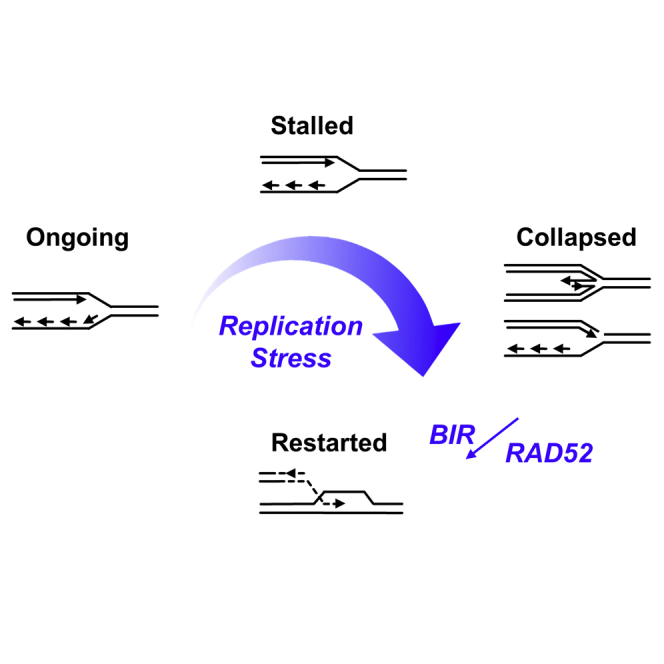
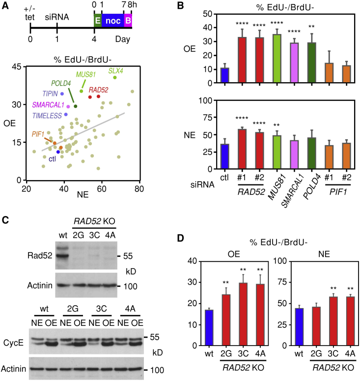
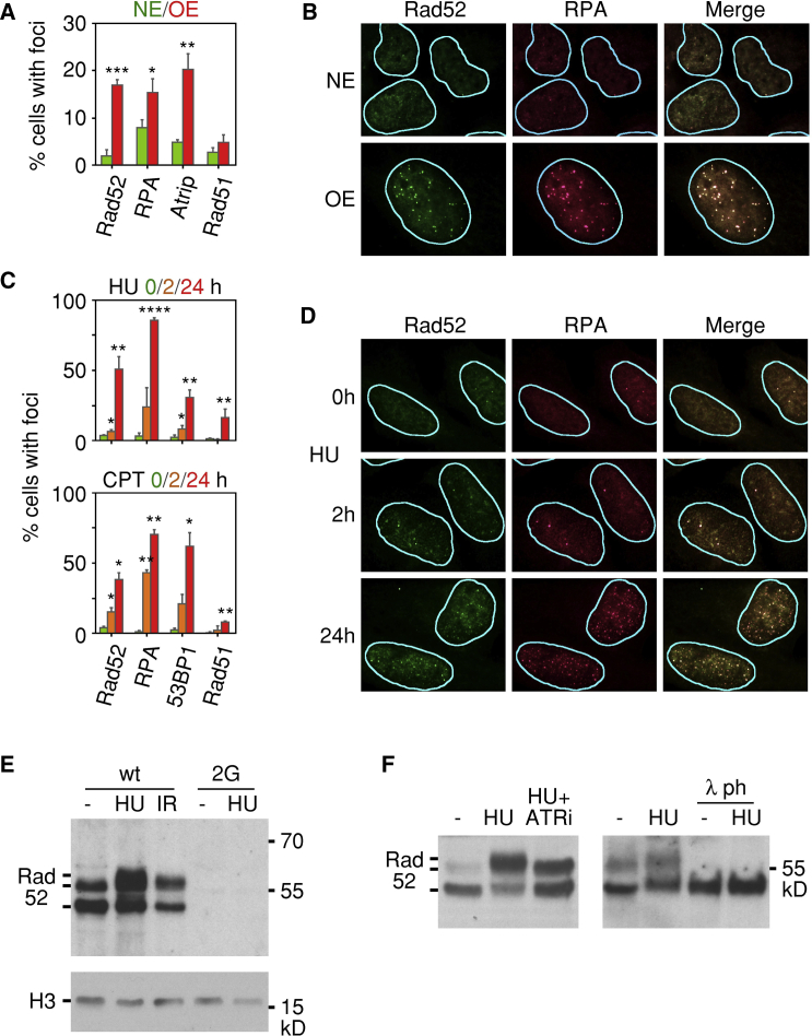
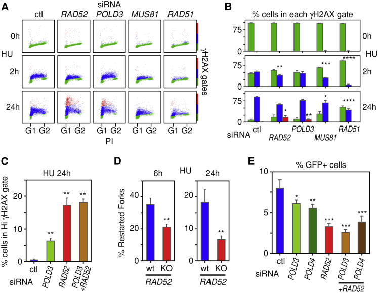
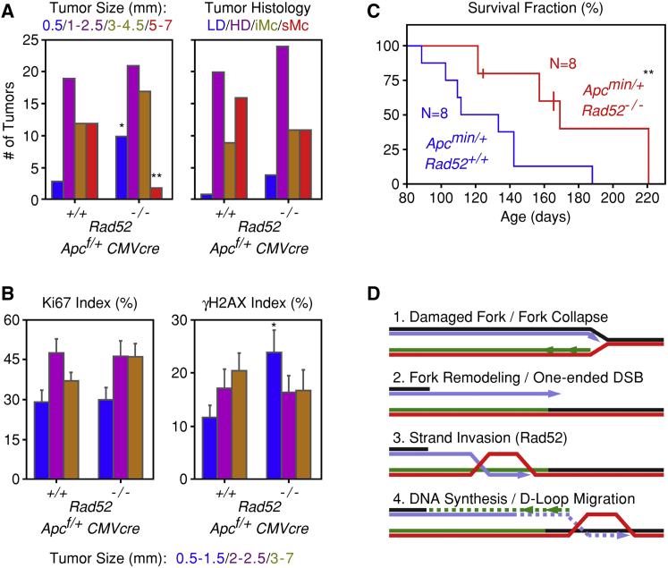
Human cancers are characterized by the presence of oncogene-induced DNA replication stress (DRS), making them dependent on repair pathways such as break-induced replication (BIR) for damaged DNA replication forks. To better understand BIR, we performed a targeted siRNA screen for genes whose depletion inhibited G1 to S phase progression when oncogenic cyclin E was overexpressed. RAD52, a gene dispensable for normal development in mice, was among the top hits. In cells in which fork collapse was induced by oncogenes or chemicals, the Rad52 protein localized to DRS foci. Depletion of Rad52 by siRNA or knockout of the gene by CRISPR/Cas9 compromised restart of collapsed forks and led to DNA damage in cells experiencing DRS. Furthermore, in cancer-prone, heterozygous APC mutant mice, homozygous deletion of the Rad52 gene suppressed tumor growth and prolonged lifespan. We therefore propose that mammalian RAD52 facilitates repair of collapsed DNA replication forks in cancer cells.

Keywords: DNA recombination; DNA replication stress; RAD52; break-induced replication; cancer.


Figures

None
Graphical abstract
Figure 1
Figure 1
RAD52 Facilitates S Phase Entry in Cells with Oncogene-Induced DRS (A) U2OS cells overexpressing cyclin E in a tetracycline (tet)-dependent manner were seeded on plates either in the presence (normal levels of cyclin E, NE) or absence (cyclin E overexpression, OE) of tet. The next day the cells were transfected with siRNA; after 3 days, they were pulse labeled with EdU for 1 hr, and 6 hr later they were pulse labeled with BrdU for 1 hr. Nocodazole (noc) was added between the EdU and BrdU pulses to prevent mitotic cells from proceeding into G1. The percentages of EdU−/BrdU− OE and NE cells were determined by flow cytometry and plotted. Selected siRNAs are indicated: ctl, control; E, EdU; B, BrdU. (B) Means and standard deviations of EdU−/BrdU− percentages of cells transfected with the indicated siRNAs. Two different siRNAs were used to target RAD52 and PIF1. In this and all other figures, one, two, three, and four asterisks denote statistical significance levels of p < 0.05, p < 0.01, p < 0.001, and p < 0.0001, respectively, and relevant statistical parameters are listed in Table S2. (C) CRISPR/Cas9-mediated inactivation of the RAD52 gene in three different knockout (KO) clones of U2OS cells inducibly overexpressing cyclin E. Lack of Rad52 protein expression (top) and robust cyclin E induction (bottom) in the three clones were documented by immunoblot analysis. (D) CRISPR/Cas9-mediated inactivation of the RAD52 gene compromises entry into S phase preferentially in cells overexpressing cyclin E (OE) as compared to cells expressing normal cyclin E (NE) levels. Means and standard deviations of the percentages of EdU−/BrdU− cells were determined using the experimental design shown in (A).
Figure 2
Figure 2
Rad52 Is Recruited to Sites of DRS (A) Means and standard deviations of the percentages of cells displaying Rad52, RPA, Atrip, or Rad51 foci in the presence of normal (NE) or high (OE) levels of cyclin E. The results are derived from three independent experiments. (B) Representative immunofluorescence images showing colocalization of Rad52 and RPA foci in cells overexpressing cyclin E (OE). (C) Means and standard deviations of the percentages of cells displaying Rad52, RPA, 53BP1, or Rad51 foci following treatment with HU or CPT for 0, 2, or 24 hr. The results are derived from three independent experiments. (D) Representative immunofluorescence images showing colocalization of Rad52 and RPA foci in cells treated with HU for 24 hr. (E) Posttranslational modifications of chromatin-bound Rad52 in cells treated with hydroxyurea (HU) for 24 hr or exposed to ionizing radiation (IR). U2OS parental cells (WT) and clone 2G with both alleles of RAD52 inactivated were cultured in the presence of tet to maintain normal levels of cyclin E. The bands corresponding to Rad52 are indicated. (F) ATR dependence of HU-induced posttranslational modification of chromatin-bound Rad52. U2OS parental cells were cultured in the presence of HU with or without an ATR inhibitor (ATRi) for 24 hr before being harvested. Tet was present in the media to maintain normal levels of cyclin E. Where indicated, the chromatin extracts were treated with lambda phosphatase (λ ph).
Figure 3
Figure 3
Rad52 Is Required for Fork Restart after Prolonged Exposure of Cells to HU (A) Rad52 and PolD3 regulate the cellular response to DRS, as ascertained by monitoring histone H2AX phosphorylation (γH2AX) in cells treated with HU for 2 or 24 hr. γH2AX levels were monitored by flow cytometry of cells treated with control (ctl) siRNA or siRNAs targeting RAD52, POLD3, MUS81, or RAD51. PI, propidium iodide. (B) Means and standard deviations of the percentages of cells assigned to the H2AX phosphorylation gates shown in (A), as determined from three independent experiments. Green, blue, and red indicate background, modest, and high H2AX phosphorylation, respectively. (C) Rad52 and PolD3 regulate the cellular response to DRS epistatically. siRNA-transfected cells were exposed to HU for 24 hr. Means and standard deviations of the percentages of cells assigned to the high (Hi) H2AX phosphorylation gate were derived from two independent experiments. (D) Fork restart after prolonged exposure of cells to HU is dependent on Rad52. U2OS parental cells (WT) and clone 2G with both alleles of RAD52 inactivated were cultured in the presence of tet to maintain normal levels of cyclin E. The cells were pulse labeled with CldU for 1 hr, then exposed to HU and a Cdc7 inhibitor for 6 or 24 hr, and finally released into media containing IdU and the Cdc7 inhibitor for 1 hr to allow fork restart. Means and standard deviations of the percentages of restarted forks were derived from three independent DNA fiber experiments. (E) Effect of depletion of PolD3, PolD4, or Rad52 on repair of DNA DSBs by BIR. Means and standard deviations of the percentages of GFP-positive cells were derived from three independent experiments.
Figure 4
Figure 4
Rad52 Deficiency Restrains Tumor Growth and Prolongs Survival of Mice with APC Mutations (A) Comparison of tumors present in the intestines of Rad52+/+;Apcf/+;CMVcre (N = 6) and Rad52−/−;Apcf/+;CMVcre (N = 6) mice. Tumors were stratified according to size (in mm) or according to histopathological criteria: LD, low-grade dysplasia; HD, high-grade dysplasia; iMc, intramucosal; and sMc, submucosal. (B) Proliferation (Ki67) and DNA damage (γH2AX) indices of the tumors present in the intestines of the Rad52+/+;Apcf/+;CMVcre and Rad52−/−;Apcf/+;CMVcre mice. Means and standard deviations of the indices were calculated after stratifying the tumors into three groups according to size. (C) Survival fractions of Rad52+/+;Apcmin/+ (N = 8) and Rad52−/−;Apcmin/+ (N = 8) mice. Three of the Rad52−/−;Apcmin/+ mice, indicated by vertical lines in the graph, had not died at the time the data were recorded and were considered censored for the statistical analysis. (D) Proposed model for the role of Rad52 in BIR.

Comment in

  • Stressing Out About RAD52.
    Ciccia A, Symington LS. Ciccia A, et al. Mol Cell. 2016 Dec 15;64(6):1017-1019. doi: 10.1016/j.molcel.2016.11.036. Mol Cell. 2016. PMID: 27984741

References

    1. Anand R.P., Lovett S.T., Haber J.E. Break-induced DNA replication. Cold Spring Harb. Perspect. Biol. 2013;5:a010397. - PMC - PubMed
    1. Arlt M.F., Wilson T.E., Glover T.W. Replication stress and mechanisms of CNV formation. Curr. Opin. Genet. Dev. 2012;22:204–210. - PMC - PubMed
    1. Bartkova J., Horejsí Z., Koed K., Krämer A., Tort F., Zieger K., Guldberg P., Sehested M., Nesland J.M., Lukas C. DNA damage response as a candidate anti-cancer barrier in early human tumorigenesis. Nature. 2005;434:864–870. - PubMed
    1. Bartkova J., Rezaei N., Liontos M., Karakaidos P., Kletsas D., Issaeva N., Vassiliou L.V., Kolettas E., Niforou K., Zoumpourlis V.C. Oncogene-induced senescence is part of the tumorigenesis barrier imposed by DNA damage checkpoints. Nature. 2006;444:633–637. - PubMed
    1. Bétous R., Mason A.C., Rambo R.P., Bansbach C.E., Badu-Nkansah A., Sirbu B.M., Eichman B.F., Cortez D. SMARCAL1 catalyzes fork regression and Holliday junction migration to maintain genome stability during DNA replication. Genes Dev. 2012;26:151–162. - PMC - PubMed

Publication types

MeSH terms