Review
. 2017 Feb:42:84-92.
doi: 10.1016/j.conb.2016.11.010. Epub 2016 Dec 13.

Timing temporal transitions during brain development

Affiliations
Review

Timing temporal transitions during brain development

Anthony M Rossi et al. Curr Opin Neurobiol. 2017 Feb.

Abstract

During development a limited number of progenitors generate diverse cell types that comprise the nervous system. Neuronal diversity, which arises largely at the level of neural stem cells, is critical for brain function. Often these cells exhibit temporal patterning: they sequentially produce neurons of distinct cell fates as a consequence of intrinsic and/or extrinsic cues. Here, we review recent advances in temporal patterning during neuronal specification, focusing on conserved players and mechanisms in invertebrate and vertebrate models. These studies underscore temporal patterning as an evolutionarily conserved strategy to generate neuronal diversity. Understanding the general principles governing temporal patterning and the molecular players involved will improve our ability to direct neural progenitors towards specific neuronal fates for brain repair.


Conflict of interest statement

Conflict of interest

Nothing declared

Conflict of interest

The authors declare that they have no conflicts of interest.

Figures
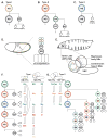
Figure 1
Figure 1. Neuroblast division modes and mechanisms of temporal patterning in Drosophila
(A–C) The three main division modes used during Drosophila neurogenesis. Type I is the result of self-renewing asymmetric divisions that produce a neuroblast (NB) and an intermediate precursor called a ganglion mother cell (GMC), which divides only once to produce two cells. Type II is a self-renewing division that produces a NB and intermediate neural progenitor (INP), which then asymmetrically divides multiple times to give rise to GMCs that only divide once. Finally, type 0 is the result of self-renewing asymmetric divisions that produce a neuroblast (NB) and a single neuron. (D) Neuroblasts delaminate from the embryonic, single-layered epithelium. Neuroblasts then divide asymmetrically (type I mode depicted here) throughout development. After each new GMC divides, earlier born neurons are displaced deeper into the embryo, resulting in a laminar organization with the earliest born neurons (green layer) occupying the deepest layer and the youngest neurons (blue layer) positioned closest to the surface. (E) The larval brain is located in the anterior of the animal and will grow throughout development as new neurons are added from NBs. At this stage, the brain can be split into three main sections, the optic lobe, the central brain, and the ventral nerve cord (VNC), each containing populations of NBs. Depicted here, optic lobe NBs from the outer proliferation center (OPC), which will give rise to medulla neurons, central brain type II NBs, which give rise to a diverse set of central brain neurons, and mushroom body NBs, which produce the intrinsic neurons of the mushroom body. (F) Embryonic and larval OPC NBs transition through three independent temporal series to dictate temporal fate. The result of this type of patterning is the ordered birth of different neuron types from single progenitors. In the embryonic VNC, most NBs transition through Hb → Kr → Pdm → Cas → Grh. In the optic lobe, NBs transition through either Hth → Klu → Ey → Slp → D → Tll or Dll → Ey → Slp → D. Temporal transcription factors do not specify a single cell fate but rather define early to late temporal identities in multiple lineages. Importantly, these factors can regulate each other, offering a model for progression through the temporal series. (G) In the type II NBs of the central brain, an additional layer of temporal patterning is added in INPs. Neuroblasts transition through D+Cas → Svp → unknown factor, while INPs progress through D → Grh → Ey. Combinatorial temporal pattering allows for each temporal transcription factor in the NB to give rise to multiple neuron types without extending the NB lineage.
Figure 2
Figure 2. Temporal patterning in the vertebrate retina and cortex
(A) There are seven main cell types in the adult retina: ganglion cells, horizontal cells, cone cells, amacrine cells, bipolar cells, rod cells and Müller glia. Although the birth order of retinal cell types overlaps highly, and individual retinal progenitor cells (RPCs) produce variable clone sizes, cone cells, horizontal cells, amacrine cells, and retinal ganglion cells are born early (green cells) whereas rod cells, bipolar cells and Muller glial cells are born late (purple cells). Early-fate specifying transcription factors are biased to be expressed early, perhaps as a result of the temporal transcription factor Ikzf1, which is expressed in RPCs early in development. In contrast, late-fate specifying transcription factors are biased to be expressed late, perhaps as a result of RPCs expressing the temporal transcription factor Casz1 late in development. The cross-regulatory interaction between Ikzf1 and Casz1 is reminiscent of the interaction between temporal transcription factors in Drosophila. (B) Initially, RPCs tend to undergo PP divisions to expand the progenitor population, before switching to PD and DD divisions (See Box 2). Although progenitors are capable of producing any cell type in any temporal window, they are biased to produce specific fates as they age (See Box 2). Interestingly, cell fate correlates with division mode: retinal ganglion cells are born from PD divisions, amacrine cells arise from both PD and DD divisions, whereas horizontal, bipolar, rod, and cone cells tend to be born from DD divisions. (C) The vertebrate cortex is built in an inside-out fashion, where early born cells reside in the deepest layer and later born cells migrate past their earlier born siblings. Neuroepithelial cells initially undergo PP division to expand the progenitor population before giving rise to radial glial cells, which are multipotent cortical progenitors. Radial glia divide asymmetrically to give rise to cells with limited proliferative potential called intermediate precursor cells (IPCs). Early in development, cortical progenitors express the transcription factor Ikzf1, a candidate temporal transcription factor associated with early-born neuronal fates, which is also expressed in early progenitors in the mammalian retina.

References

    1. Azzarelli R, Hardwick LJA, Philpott A. Emergence of neuronal diversity from patterning of telencephalic progenitors. Wiley Interdiscip Rev Dev Biol. 2015;4:197–214. - PubMed
    1. Urbach R, Technau GM. Neuroblast formation and patterning during early brain development in Drosophila. Bioessays. 2004;26:739–51. - PubMed
    1. Alfano C, Studer M. Neocortical arealization: Evolution, mechanisms, and open questions. Dev Neurobiol. 2013;73:411–447. - PubMed
    1. Dasen JS, Jessell TM. Hox networks and the origins of motor neuron diversity. Curr Top Dev Biol. 2009;88:169–200. - PubMed
    1. Kohwi M, Doe CQ. Temporal fate specification and neural progenitor competence during development. Nat Rev Neurosci. 2013;14:823–838. - PMC - PubMed