Review
. 2017 Apr;27(4):241-254.
doi: 10.1016/j.tcb.2016.10.005. Epub 2016 Dec 13.

Mechanical Communication at the Immunological Synapse

Affiliations
Review

Mechanical Communication at the Immunological Synapse

Roshni Basu et al. Trends Cell Biol. 2017 Apr.

Abstract

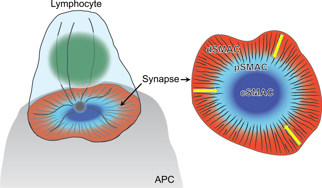
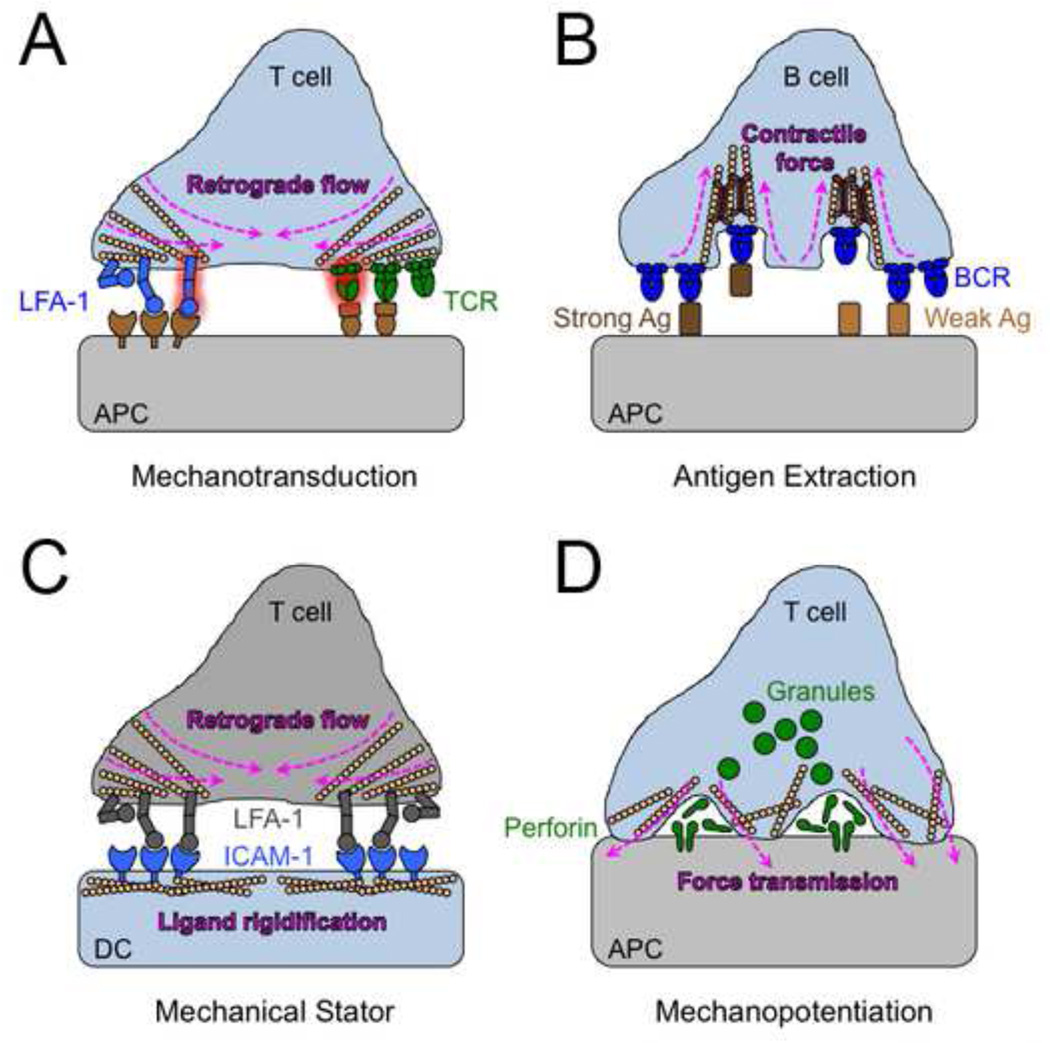
T and B lymphocytes communicate by forming immunological synapses with antigen-presenting target cells. These highly dynamic contacts are characterized by continuous cytoskeletal remodeling events, which not only structure the interface but also exert a considerable amount of mechanical force. In recent years, it has become increasingly clear that synaptic forces influence information transfer both into and out of the lymphocyte. Here, we review our current understanding of synapse mechanics, focusing on its role as an avenue for intercellular communication.

Keywords: B cell; T cell; immunology; mechanobiology; signal transduction.


Figures

Figure 1
Figure 1. Schematic diagram of the mature IS
A side view is shown to the left and an en face view to the right. The lymphocyte nucleus is colored green, with F-actin and microtubules depicted as black and gray lines, respectively. Yellow arrows on the right indicate the direction of retrograde F-actin flow.
Figure 2
Figure 2. Mechanical information transfer at the IS
(A) Pulling forces derived from retrograde F-actin flow within the lymphocyte promote the mechanical activation of LFA-1 and the TCR. (B) Contractile forces driven by actin and myosin generate membrane invaginations that facilitate the discrimination between strong and weak antigens by the BCR. (C) DCs use their cortical cytoskeleton to constrain the diffusion of ICAM-1, thereby promoting mechanical activation of LFA-1 on the T cell side of the IS. (D) CTLs use mechanical force to strain the surface of the target cell, which boosts killing responses by potentiating the pore forming activity of perforin.

References

    1. Alon R, Feigelson SW, Manevich E, Rose DM, Schmitz J, Overby DR, Ginsberg MH. Alpha4beta1-dependent adhesion strengthening under mechanical strain is regulated by paxillin association with the alpha4-cytoplasmic domain. J Cell Biol. 2005;171:1073–1084. - PMC - PubMed
    1. Astrof NS, Salas A, Shimaoka M, Chen J, Springer TA. Importance of force linkage in mechanochemistry of adhesion receptors. Biochemistry. 2006;45:15020–15028. - PMC - PubMed
    1. Bashour KT, Gondarenko A, Chen H, Shen K, Liu X, Huse M, Kam LC. CD28 and CD3 have complementary roles in T-cell traction forces. Proc Natl Acad Sci U S A. 2014;111:2241–2246. - PMC - PubMed
    1. Basu R, Whitlock BM, Husson J, Le Floc'h A, Jin W, Oyler-Yaniv A, Huse M. Cytotoxic T Cells Use Mechanical Force to Potentiate Target Cell Killing. Cell. 2016 - PMC - PubMed
    1. Benvenuti F. The Dendritic Cell Synapse: A Life Dedicated to T Cell Activation. Front Immunol. 2016;7:70. - PMC - PubMed

Publication types