. 2017 Feb;30(2):95-103.
doi: 10.1093/protein/gzw067. Epub 2016 Dec 15.

Development of a cancer-marker activated enzymatic switch from the herpes simplex virus thymidine kinase

Affiliations

Development of a cancer-marker activated enzymatic switch from the herpes simplex virus thymidine kinase

Nirav Y Shelat et al. Protein Eng Des Sel. 2017 Feb.

Abstract

Discovery of new cancer biomarkers and advances in targeted gene delivery mechanisms have made gene-directed enzyme prodrug therapy (GDEPT) an attractive method for treating cancer. Recent focus has been placed on increasing target specificity of gene delivery systems and reducing toxicity in non-cancer cells in order to make GDEPT viable. To help address this challenge, we have developed an enzymatic switch that confers higher prodrug toxicity in the presence of a cancer marker. The enzymatic switch was derived from the herpes simplex virus thymidine kinase (HSV-TK) fused to the CH1 domain of the p300 protein. The CH1 domain binds to the C-terminal transactivation domain (C-TAD) of the cancer marker hypoxia inducible factor 1α. The switch was developed using a directed evolution approach that evaluated a large library of HSV-TK/CH1 fusions using a negative selection for azidothymidine (AZT) toxicity and a positive selection for dT phosphorylation. The identified switch, dubbed TICKLE (Trigger-Induced Cell-Killing Lethal-Enzyme), confers a 4-fold increase in AZT toxicity in the presence of C-TAD. The broad substrate specificity exhibited by HSV-TK makes TICKLE an appealing prospect for testing in medical imaging and cancer therapy, while establishing a foundation for further engineering of nucleoside kinase protein switches.

Keywords: HSV; directed evolution; herpes simplex virus thymidine kinase; protein switch; thymidine kinase.


Figures

Fig. 1
Fig. 1
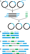
Construction of libraries. (A) Libraries with the CH1 domain inserted into HSV-TK were created using inverse PCR in which the plasmid containing hsv-tk was linearized at specific codons within the gene. The linearized plasmid was ligated with CH1 DNA to create the circularized plasmid library with the CH1 inserted into HSV-TK at different sites. Various linkers of different composition and length were added to the CH1 DNA by PCR before the ligation step to generate the libraries: (B) pNYS01, (C) pNYS02 and (D) pNYS03. Linkers shown are illustrative of the diversity.
Fig. 2
Fig. 2
Genetic selections. (A) The negative selection in E. coli KY895 identifies variants that lack kinase activity in the absence of the C-TAD of HIF1-α. Variants that possess activity in this condition produce toxic AZT-MP, while variants lacking kinase activity do not and thus survive. (B) The positive selection, also done in E. coli KY895, selects for variants with the ability to phosphorylate dT to dTMP in the presence of the C-TAD. Cells that are unable metabolize the necessary dTMP are killed.
Fig. 3
Fig. 3
TICKLE switch. (A) Sequence of TICKLE. TICKLE comprised a CH1 domain insert in between the 150th and 151st residues of HSV-TK. The N-terminus of the CH1 is connected to HSV-TK by a Gly-Gly-Ser linker and the C-terminus is connected by a Ser-Thr-Thr linker. (B) Structure models of HSV-TK (PDB #1KIM (Champness et al., 1998)) adjacent to CH1 with the C-TAD of HIF1-α bound (PDB #1LE3 (Freedman et al., 2002)). Space-filled spheres indicate the sites of fusion (i.e. insertion) of CH1 into HSV-TK. The insertion site in HSV-TK (between residues 150 and 151) is within an unstructured loop (residues 148–153) in the crystal structure, thus residues 147 and 154 are indicated instead. Thymidine (dT) is shown in the active site of HSV-TK.
Fig. 4
Fig. 4
AZT toxicity assay. (A) C-TAD coexpression increases the AZT toxicity of E. coli KY895 cells expressing TICKLE. An equal number of cells expressing TICKLE + GST or TICKLE + C-TAD-GST were spotted on negative selection plates containing 10 μg/ml AZT and incubated 18 hours at 37°C. The different spots represent a series of 1:2 serial dilutions of the culture, with the highest cell concentration at the right. (B) Escherichia coli KY895 cells expressing the indicated proteins were challenged to grow in liquid media for 6 hours in the presence of different concentrations of AZT. The relative OD600 normalizes the OD600 to that in the absence of AZT. Circles, TICKLE + C-TAD-GST; squares, TICKLE + GST; triangles, HSV-TK (positive control) and diamonds, inactive HSV-TK (negative control). (C) Circles, TICKLE (T244S) + C-TAD-GST; squares, TICKLE (T244S) + GST; triangles, TICKLE (S243G, T244G, T245S) + GST; diamonds, TICKLE (S243G, T244G, T245S) + C-TAD-GST. Error bars represent the standard deviation (n = 3).
Fig. 5
Fig. 5
dT growth assay. (A) Escherichia coli NS01 is a conditional lethal strain that requires rhamnose for growth due to a deficiency in dTMP production. One thousand CFU cells were plated on tryptone media plates with 25 μg/ml kanamycin and containing 0.2% glucose (Glu) (top; non-permissive condition) or 0.2% rhamnose (Rha) (bottom; permissive condition) and incubated 18 hours at 37°C. (B) Escherichia coli NS01 expressing the following proteins were grown in non-permissive liquid media for 8 hours in the presence of different concentrations of dT. Circles, TICKLE + C-TAD-GST; squares, TICKLE + GST; triangles, inactive HSV-TK (negative control) and diamonds, wild-type HSV-TK (positive control). The relative OD600 was normalized to the OD600 of the positive control at 50 μg/ml dT. Error bars represent the standard deviation (n = 3). Points that appear to be without error bars result from errors that are smaller than the size of the symbol.
Fig. 6
Fig. 6
Relative accumulation of TICKLE as a function of C-TAD coexpression. A western blot using anti-HSV-TK antibodies was performed on an SDS-PAGE gel of the soluble fraction from an equal number of KY895 cells expressing the indicated proteins. Lane 1, HSV-TK; Lane 3, TICKLE + C-TAD-GST; Lane 4, TICKLE + GST; Lane 5, no heterologous protein expressed. Lane 2 is the molecular weight marker.

References

    1. Aghi M., Kramm C.M., Chou T.C., Breakefield X.O. and Chiocca E.A. (1998) J. Natl. Cancer Inst., 90, 370–380. - PubMed
    1. Ahmad S.I., Kirk S.H. and Eisenstark A. (1998) Annu. Rev. Microbiol., 52, 591–625. - PubMed
    1. Bai L. and Merchant J.L. (2007) FEBS Lett., 581, 5904–5910. - PMC - PubMed
    1. Black M.E., Newcomb T.G., Wilson H.M. and Loeb L.A. (1996) Proc. Natl. Acad. Sci. USA, 93, 3525–3529. - PMC - PubMed
    1. Bosley A.D. and Ostermeier M. (2005) Biomol. Eng., 22, 57–61. - PubMed

MeSH terms