

Histological Underpinnings of Grey Matter Changes in Fibromyalgia Investigated Using Multimodal Brain Imaging
- PMID: 27986927
- PMCID: PMC6596849
- DOI: 10.1523/JNEUROSCI.2619-16.2016
Histological Underpinnings of Grey Matter Changes in Fibromyalgia Investigated Using Multimodal Brain Imaging
Abstract
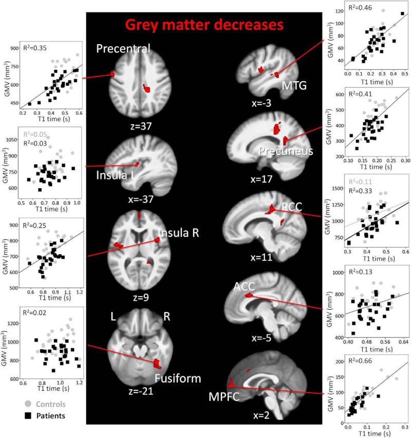
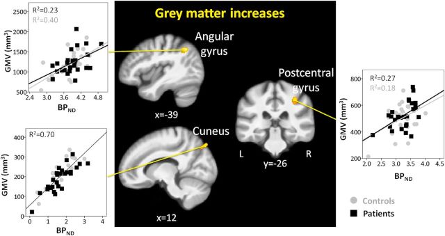
Chronic pain patients present with cortical gray matter alterations, observed with anatomical magnetic resonance (MR) imaging. Reduced regional gray matter volumes are often interpreted to reflect neurodegeneration, but studies investigating the cellular origin of gray matter changes are lacking. We used multimodal imaging to compare 26 postmenopausal women with fibromyalgia with 25 healthy controls (age range: 50-75 years) to test whether regional gray matter volume decreases in chronic pain are associated with compromised neuronal integrity. Regional gray matter decreases were largely explained by T1 relaxation times in gray matter, a surrogate measure of water content, and not to any substantial degree by GABAA receptor concentration, an indirect marker of neuronal integrity measured with [18F] flumazenil PET. In addition, the MR spectroscopy marker of neuronal viability, N-acetylaspartate, did not differ between patients and controls. These findings suggest that decreased gray matter volumes are not explained by compromised neuronal integrity. Alternatively, a decrease in neuronal matter could be compensated for by an upregulation of GABAA receptors. The relation between regional gray matter and T1 relaxation times suggests decreased tissue water content underlying regional gray matter decreases. In contrast, regional gray matter increases were explained by GABAA receptor concentration in addition to T1 relaxation times, indicating perhaps increased neuronal matter or GABAA receptor upregulation and inflammatory edema. By providing information on the histological origins of cerebral gray matter alterations in fibromyalgia, this study advances the understanding of the neurobiology of chronic widespread pain.
Significance statement: Regional gray matter alterations in chronic pain, as detected with voxel-based morphometry of anatomical magnetic resonance images, are commonly interpreted to reflect neurodegeneration, but this assumption has not been tested. We found decreased gray matter in fibromyalgia to be associated with T1 relaxation times, a surrogate marker of water content, but not with GABAA receptor concentration, a surrogate of neuronal integrity. In contrast, regional gray matter increases were partly explained by GABAA receptor concentration, indicating some form of neuronal plasticity. The study emphasizes that voxel-based morphometry is an exploratory measure, demonstrating the need to investigate the histological origin of gray matter alterations for every distinct clinical entity, and advances the understanding of the neurobiology of chronic (widespread) pain.
Keywords: chronic pain; flumazenil PET; grey matter; neurodegeneration; voxel-based morphometry.
Copyright © 2017 the authors 0270-6474/17/371091-12$15.00/0.
Figures
References
-
- Bennett R. (2005) The Fibromyalgia Impact Questionnaire (FIQ): a review of its development, current version, operating characteristics and uses. Clin Exp Rheumatol 23:S154–S162. - PubMed
Publication types
MeSH terms
Substances
Grants and funding
LinkOut - more resources
Full Text Sources
Other Literature Sources
Medical