. 2017;9(2):193-202.
doi: 10.1159/000451007. Epub 2016 Dec 17.

Pigs with Severe Combined Immunodeficiency Are Impaired in Controlling Influenza A Virus Infection

Affiliations

Pigs with Severe Combined Immunodeficiency Are Impaired in Controlling Influenza A Virus Infection

Daniela S Rajao et al. J Innate Immun. 2017.

Abstract

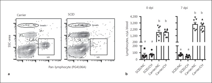
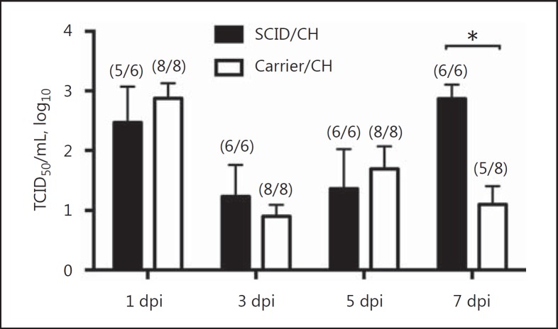
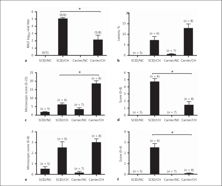
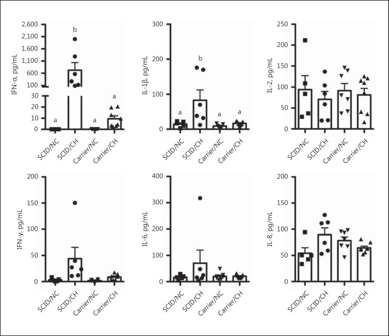
Influenza A viruses (IAV) infect many host species, including humans and pigs. Severe combined immunodeficiency (SCID) is a condition characterized by a deficiency of T, B, and/or natural killer (NK) cells. Animal models of SCID have great value for biomedical research. Here, we evaluated the pathogenesis and the innate immune response to the 2009 H1N1 pandemic IAV (H1N1pdm09) using a recently identified line of naturally occurring SCID pigs deficient in T and B lymphocytes that still have functional NK cells. SCID pigs challenged with H1N1pdm09 showed milder lung pathology compared to the non-SCID heterozygous carrier pigs. Viral titers in the lungs and nasal swabs of challenged SCID pigs were significantly higher than in carrier pigs 7 days postinfection, despite higher levels of IL-1β and IFN-α in the lungs of SCID pigs. The lower levels of pulmonary pathology were associated with the T and B cell absence in response to infection. The higher viral titers, prolonged shedding, and delayed viral clearance indicated that innate immunity was insufficient for controlling IAV in pigs. This recently identified line of SCID pigs provides a valuable model to understand the immune mechanisms associated with influenza protection and recovery in a natural host.


Figures

Fig. 1
Fig. 1
Enumeration of peripheral lymphocytes by flow cytometry confirmed the SCID phenotype. Peripheral blood was collected and the number of circulating lymphocytes labeled with a pan-lymphocyte antibody (clone PG106A) as described in the Materials and Methods. a Representative flow plots of lymphocyte populations (PG106A+) in carrier and SCID pigs. Beads were used to enumerate the number of lymphocytes (LO). b The number of lymphocytes in the indicated groups on the day of challenge (0 dpi) and necropsy (7 dpi). Each individual symbol indicates the value for a pig in that respective treatment group, with mean and SEM also indicated. Bars with different letters indicate statistical differences (p ≤ 0.05). CH, challenged; NC, nonchallenged control.
Fig. 2
Fig. 2
Virus titers in nasal swabs at 1, 3, 5, and 7 dpi of pigs with SCID or carrier control pigs challenged (CH) with A/California/04/2009. The numbers of infected pigs/total number of pigs are indicated in parentheses. Results are shown as means and SEM of log10 titers. Significant differences (* p ≤ 0.05) within each dpi are indicated.
Fig. 3
Fig. 3
Influenza effects on the lower respiratory tract differ in SCID pigs. Virus titers in BALF at 7 dpi (a), macroscopic pneumonia (b), lung microscopic pathology (c), IAV antigen detection scores in lungs (d), trachea microscopic pathology (e), and IAV antigen detection scores in trachea (f) in pigs with SCID or carrier control pigs challenged (CH) with A/California/04/2009, and nonchallenged controls (NC). The numbers of pigs per group or numbers of infected pigs/total number of pigs are indicated in parentheses. The results are shown as means and SEM. Significant differences (* p ≤ 0.05) between challenged groups are indicated.
Fig. 4
Fig. 4
Levels of IFN-α, IL-1β, IL-2, IFN-γ, IL-6, and IL-8 in the lungs of pigs with SCID or carrier control pigs challenged (CH) with A/California/04/2009, and nonchallenged controls (NC). Lung lavage was collected on day 7 following challenge, and the amount of each indicated cytokine was measured by multiplex ELISA. Each individual symbol indicates the value for a pig in that respective treatment group, with the mean and SEM also indicated. Bars with different letters indicate statistical differences (p ≤ 0.05).
Fig. 5
Fig. 5
Serum antibody detection at 7 dpi in pigs with SCID or carrier control pigs challenged (CH) with A/California/04/2009, and nonchallenged controls (NC) by blocking ELISA. Values are shown as mean S/N ratios and SEM, with lower S/N ratios indicative of the presence of IAV-specific antibodies. The cut-off value for a negative sample is indicated by the dotted line, and the maximum value for a sample to be considered positive is indicated by the solid line (positive threshold) with a suspect range between the two. The number of pigs per group is indicated in parentheses. Significant differences (* p ≤ 0.05) between groups are indicated.

Similar articles

Cited by

References

    1. Perryman LE. Molecular pathology of severe combined immunodeficiency in mice, horses, and dogs. Vet Pathol. 2004;41:95–100. - PubMed
    1. Bauer TR, Jr, Adler RL, Hickstein DD. Potential large animal models for gene therapy of human genetic diseases of immune and blood cell systems. ILAR J. 2009;50:168–186. - PMC - PubMed
    1. Suzuki S, Iwamoto M, Saito Y, Fuchimoto D, Sembon S, Suzuki M, Mikawa S, Hashimoto M, Aoki Y, Najima Y, Takagi S, Suzuki N, Suzuki E, Kubo M, Mimuro J, Kashiwakura Y, Madoiwa S, Sakata Y, Perry AC, Ishikawa F, Onishi A. Il2rg gene-targeted severe combined immunodeficiency pigs. Cell Stem Cell. 2012;10:753–758. - PubMed
    1. Watanabe M, Nakano K, Matsunari H, Matsuda T, Maehara M, Kanai T, Kobayashi M, Matsumura Y, Sakai R, Kuramoto M, Hayashida G, Asano Y, Takayanagi S, Arai Y, Umeyama K, Nagaya M, Hanazono Y, Nagashima H. Generation of interleukin-2 receptor gamma gene knockout pigs from somatic cells genetically modified by zinc finger nuclease-encoding mRNA. PLoS One. 2013;8:e76478. - PMC - PubMed
    1. Lee K, Kwon DN, Ezashi T, Choi YJ, Park C, Ericsson AC, Brown AN, Samuel MS, Park KW, Walters EM, Kim DY, Kim JH, Franklin CL, Murphy CN, Roberts RM, Prather RS, Kim JH. Engraftment of human iPS cells and allogeneic porcine cells into pigs with inactivated RAG2 and accompanying severe combined immunodeficiency. Proc Natl Acad Sci USA. 2014;111:7260–7265. - PMC - PubMed

MeSH terms