

Photobiomodulation reduces drusen volume and improves visual acuity and contrast sensitivity in dry age-related macular degeneration
- PMID: 27989012
- PMCID: PMC5484346
- DOI: 10.1111/aos.13354
Photobiomodulation reduces drusen volume and improves visual acuity and contrast sensitivity in dry age-related macular degeneration
Abstract
Purpose: To evaluate the efficacy of photobiomodulation (PBM) treatment for patients with dry age-related macular degeneration (AMD).
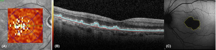
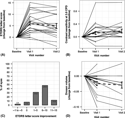
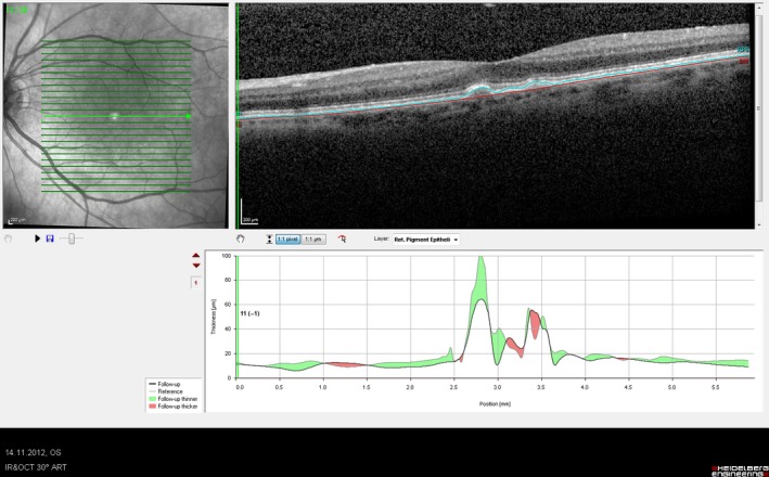
Methods: Assessments on 42 eyes with dry AMD (age related eye disease study (AREDS) 2-4) were conducted. Multiwavelength light emitting diode (LED) light comprising of yellow (590 nm), red (670 nm) and near-infrared (790 nm) bandwidths was applied to subjects' eyes for a treatment course of 3 weeks. Outcome measures were changes in best-corrected visual acuity (BCVA), contrast sensitivity (CS), drusen volume and central drusen thickness.
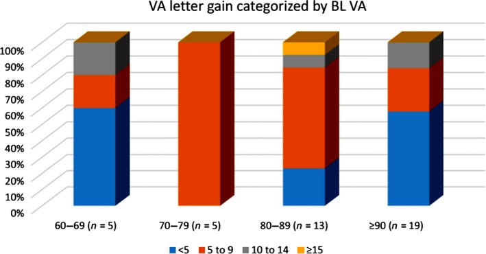
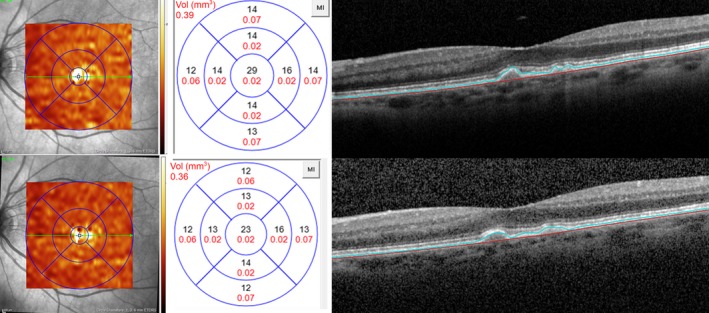
Results: Significant improvement in mean BCVA of 5.90 letters (p < 0.001) was seen on completion of the 3-week treatment and 5.14 letters (p < 0.001) after 3 months. Contrast sensitivity improved significantly (log unit improvement of 0.11 (p = 0.02) at 3 weeks and 3 months (log unit improvement of 0.16 (p = 0.02) at three cycles per degree. Drusen volume decreased by 0.024 mm3 (p < 0.001) and central drusen thickness was significantly reduced by a mean of 3.78 μm (p < 0.001), while overall central retinal thickness and retinal volume remained stable.
Conclusion: This is the first study demonstrating improvements in functional and anatomical outcomes in dry AMD subjects with PBM therapy. These findings corroborate an earlier pilot study that looked at functional outcome measures. The addition of anatomical evidence contributes to the basis for further development of a non-invasive PBM treatment for dry AMD.
Keywords: age-related macular degeneration; contrast; drusen; photobiomodulation; sensitivity; visual acuity.
© 2016 The Authors. Acta Ophthalmologica published by John Wiley & Sons Ltd on behalf of Acta Ophthalmologica Scandinavica Foundation.
Figures
Comment in
-
Photobiomodulation in dry age-related macular degeneration.Acta Ophthalmol. 2018 Feb;96(1):e92. doi: 10.1111/aos.13433. Epub 2017 Nov 22. Acta Ophthalmol. 2018. PMID: 29164805 No abstract available.
References
-
- Albarracin R, Eells J & Valter K (2011): Photobiomodulation protects the retina from light‐induced photoreceptor degeneration. Invest Ophthalmol Vis Sci 52: 3582–3592. - PubMed
-
- Alten F & Eter N (2015): Current knowledge on reticular pseudodrusen in age‐related macular degeneration. Br J Ophthalmol 99: 717–722. - PubMed
-
- Beck RW, Moke PS, Turpin AH et al. (2003): A computerized method of visual acuity testing: adaptation of the early treatment of diabetic retinopathy study testing protocol. Am J Ophthalmol 135: 194–205. - PubMed
-
- Chowers I, Tiosano L, Audo I, Grunin M & Boon CJ (2015): Adult‐onset foveomacular vitelliform dystrophy: a fresh perspective. Prog Retin Eye Res 47: 64–85. - PubMed
MeSH terms
LinkOut - more resources
Full Text Sources
Other Literature Sources
Medical
