. 2017 Jan 5;100(1):151-159.
doi: 10.1016/j.ajhg.2016.11.014. Epub 2016 Dec 15.

Mutations in MDH2, Encoding a Krebs Cycle Enzyme, Cause Early-Onset Severe Encephalopathy

Affiliations

Mutations in MDH2, Encoding a Krebs Cycle Enzyme, Cause Early-Onset Severe Encephalopathy

Samira Ait-El-Mkadem et al. Am J Hum Genet. .

Abstract

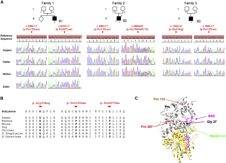
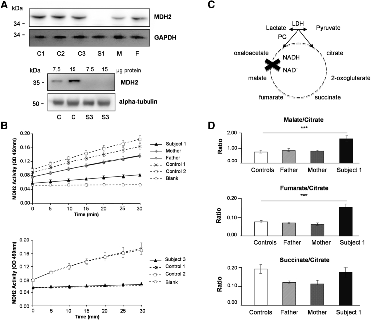
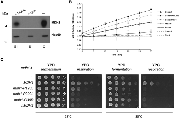
MDH2 encodes mitochondrial malate dehydrogenase (MDH), which is essential for the conversion of malate to oxaloacetate as part of the proper functioning of the Krebs cycle. We report bi-allelic pathogenic mutations in MDH2 in three unrelated subjects presenting with early-onset generalized hypotonia, psychomotor delay, refractory epilepsy, and elevated lactate in the blood and cerebrospinal fluid. Functional studies in fibroblasts from affected subjects showed both an apparently complete loss of MDH2 levels and MDH2 enzymatic activity close to null. Metabolomics analyses demonstrated a significant concomitant accumulation of the MDH substrate, malate, and fumarate, its immediate precursor in the Krebs cycle, in affected subjects' fibroblasts. Lentiviral complementation with wild-type MDH2 cDNA restored MDH2 levels and mitochondrial MDH activity. Additionally, introduction of the three missense mutations from the affected subjects into Saccharomyces cerevisiae provided functional evidence to support their pathogenicity. Disruption of the Krebs cycle is a hallmark of cancer, and MDH2 has been recently identified as a novel pheochromocytoma and paraganglioma susceptibility gene. We show that loss-of-function mutations in MDH2 are also associated with severe neurological clinical presentations in children.


Figures

Figure 1
Figure 1
MDH2 Mutations in Three Unrelated Affected Subjects (A) Pedigrees and sequence chromatograms showing variant phenotypes and segregation through the subjects’ families. (B) Cross-species conservation of the MDH2 sequence flanking the altered Gly37, Pro133, and Pro207 amino acids. (C) Three-dimensional representation of the crystal structure of human MDH2 (PDB: 2DFD, residues 24–337), shown as a homodimer with one molecule in gray and a second in yellow. Green and fuchsia sticks illustrate malate ions and NAD, respectively. The mutated residues are highlighted in black (Gly37), orange (Pro133), and red (Pro207).
Figure 2
Figure 2
MDH2 Mutations Cause Loss of MDH2 Levels and Enzymatic Activity (A) Western blot analysis with anti-MDH2 antibodies in fibroblasts from subject 1 (S1, top) and subject 3 (S3, bottom). Additional abbreviations are as follows: C1–C3, control individuals; M, mother of S1; and F, father of S1. Commercial MDH2-specific antisera at 1:250 (HPA019716, Sigma-Aldrich) or 1:1,000 (8610S, Cell Signaling Technology) were used for the upper and lower panels, respectively. GAPDH and α-tubulin were used as loading controls. (B) Representation of MDH2 activity, measured with the Mitochondrial Malate Dehydrogenase (MDH2) Activity Assay Kit (Ab119693, Abcam), in the fibroblasts from S1, his parents, and control individuals (top) and in those from S3 and control individuals (bottom). Data represent three independent experiments performed in duplicate. (C) Schematic of the Krebs cycle. (D) Metabolite ratios assessed by liquid chromatographic tandem-mass spectrometry in fibroblasts from S1 are compared with those from his parents and control individuals. Experiments were performed as previously described. Differences between fibroblast cells were analyzed by Student’s t test: ∗∗∗p < 0.001 (extremely significant).
Figure 3
Figure 3
Functional Complementation Showing the Pathogenicity of MDH2 Variants (A and B) Functional complementation of fibroblasts. The human full-length MDH2 cDNA was cloned into the pHR-SIN-CSGW dlNotI vector by PCR (Table S3). Lentiviral particles were produced in HEK293T cells. Then, fibroblasts were infected with viral supernatant expressing either MDH2 cDNA or eGFP (as a control). (A) Restoration of MDH2 levels in S1 fibroblasts transduced with wild-type MDH2 cDNA as determined by western blot with anti-MDH2 antibody. No restoration was observed when cells were transduced with GFP cDNA (C, non-transduced control fibroblasts). (B) Restoration of MDH2 activity in S1 fibroblasts transduced with wild-type MDH2 cDNA (two independent experiments performed in duplicate). No restoration was observed when cells were transduced with GFP cDNA. The MDH2 activity of non-transduced fibroblasts from parents and a control individual is also shown. (C) Functional complementation assay of the yeast mdh1Δ-null mutant. Human MDH2 cDNA was amplified with the corresponding primers (Table S3) and cloned into the centromeric expression plasmid pYX122 (constitutive promoter TPI, Addgene). The yeast wild-type MDH1 and the mdh1 mutated sequences corresponding to the three missense mutations were obtained by PCR (Table S3), and the corresponding fragments were introduced into the linearized pYX122 vector by co-transformation of the mdh1Δ strain (BY4741, Open Biosystems) and homologous recombination. Plasmids from the transformants were extracted and sequenced. These plasmids were then reintroduced into the mdh1Δ strain for testing their complementation capability. Growth of mdh1Δ (−) transformed with wild-type MDH1, mutated mdh1-P128L, mdh1-P202L, or mdh1-G30R, or wild-type hMDH2 plasmids was tested either on glucose (YPD, fermentable carbon source) or on glycerol (YPG, non-fermentable carbon source). Drop dilution growth tests were performed at 1/10 dilution steps and incubated on YPD or YPG plates for 2 days at 28°C or 35°C.

References

    1. Khurana D.S., Salganicoff L., Melvin J.J., Hobdell E.F., Valencia I., Hardison H.H., Marks H.G., Grover W.D., Legido A. Epilepsy and respiratory chain defects in children with mitochondrial encephalopathies. Neuropediatrics. 2008;39:8–13. - PubMed
    1. Sobreira N., Schiettecatte F., Valle D., Hamosh A. GeneMatcher: a matching tool for connecting investigators with an interest in the same gene. Hum. Mutat. 2015;36:928–930. - PMC - PubMed
    1. Bannwarth S., Ait-El-Mkadem S., Chaussenot A., Genin E.C., Lacas-Gervais S., Fragaki K., Berg-Alonso L., Kageyama Y., Serre V., Moore D.G. A mitochondrial origin for frontotemporal dementia and amyotrophic lateral sclerosis through CHCHD10 involvement. Brain. 2014;137:2329–2345. - PMC - PubMed
    1. Cascón A., Comino-Méndez I., Currás-Freixes M., de Cubas A.A., Contreras L., Richter S., Peitzsch M., Mancikova V., Inglada-Pérez L., Pérez-Barrios A. Whole-exome sequencing identifies MDH2 as a new familial paraganglioma gene. J. Natl. Cancer Inst. 2015;107 - PubMed
    1. Lasserre J.-P., Dautant A., Aiyar R.S., Kucharczyk R., Glatigny A., Tribouillard-Tanvier D., Rytka J., Blondel M., Skoczen N., Reynier P. Yeast as a system for modeling mitochondrial disease mechanisms and discovering therapies. Dis. Model. Mech. 2015;8:509–526. - PMC - PubMed

MeSH terms