. 2017 Jan 5;65(1):131-141.
doi: 10.1016/j.molcel.2016.10.035. Epub 2016 Dec 15.

Chromatin Constrains the Initiation and Elongation of DNA Replication

Affiliations

Chromatin Constrains the Initiation and Elongation of DNA Replication

Sujan Devbhandari et al. Mol Cell. .

Abstract

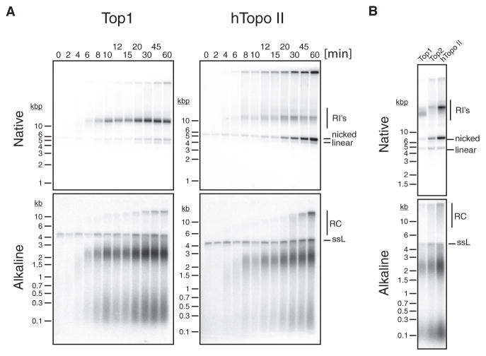
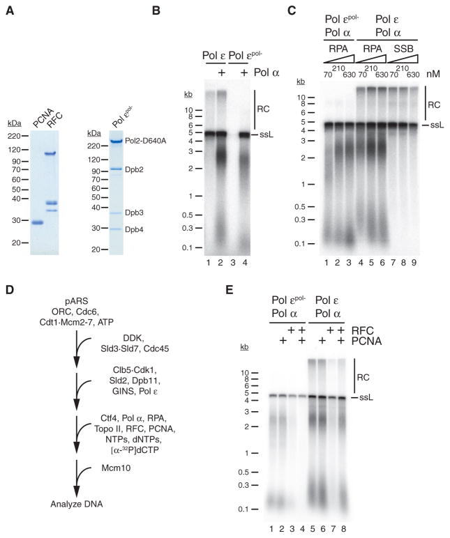
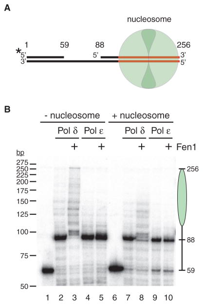
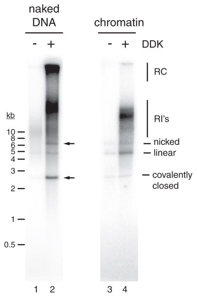
Eukaryotic chromosomal DNA is faithfully replicated in a complex series of cell-cycle-regulated events that are incompletely understood. Here we report the reconstitution of DNA replication free in solution with purified proteins from the budding yeast Saccharomyces cerevisiae. The system recapitulates regulated bidirectional origin activation; synthesis of leading and lagging strands by the three replicative DNA polymerases Pol α, Pol δ, and Pol ε; and canonical maturation of Okazaki fragments into continuous daughter strands. We uncover a dual regulatory role for chromatin during DNA replication: promoting origin dependence and determining Okazaki fragment length by restricting Pol δ progression. This system thus provides a functional platform for the detailed mechanistic analysis of eukaryotic chromosome replication.

Keywords: DNA polymerase; DNA replication; McM2-7; ORC; Okazaki fragment; chromatin; lagging strand; leading strand; replication origin.


Figures

Figure 1
Figure 1. Reconstitution of Origin Activation on Plasmid DNA Free in Solution
(A) Purified budding yeast replication initiation proteins analyzed by SDS-PAGE and Coomassie stain. (B) Reaction scheme. pARS, ARS-containing plasmid. (C) Nascent DNA products obtained with pARS1 (4.8 kb) as described in (B) were analyzed by alkaline agarose gel-electrophoresis and autoradiography. Initiation factors were individually omitted from the reaction as indicated. (D) Analysis of DNA products obtained with pARS305 (9.8 kb) as in (C). See also Figure S1.
Figure 2
Figure 2. Both Topo I and Topo II Support Replication Elongation, but Only Topo II Supports Decatenation of Plasmid Daughter Molecules
(A) Reactions were performed with pARS1 (4.8 kb) according to the scheme of Figure 1B. At indicated times after addition of Mcm10, aliquots were withdrawn from each reaction and processed for analysis by native (top) or alkaline (bottom) agarose gel-electrophoresis and autoradiography. RI’s, replication intermediates. (B) Reactions were performed with pARS1 (4.8 kb) according to the scheme of Figure 1B using Top1, Top2, or human Topo II (hTopo II) as indicated. DNA products were analyzed by native (top) or alkaline (bottom) agarose gel-electrophoresis and autoradiography. See also Figure S2.
Figure 3
Figure 3. PCNA Clamp Loading Inhibits Primer Elongation by Pol α
(A) Purified budding yeast factors. (B) Reactions were carried out with pARS1 (4.8 kb) according to the scheme in Figure 1B, using either wild-type Pol ε (lanes 1 and 2) or Pol εpo1– (lanes 3 and 4) either in the absence or presence of Pol α as indicated. DNA products were analyzed by alkaline agarose gel-electrophoresis and autoradiography. (C) Reactions were carried out with pARS1 (4.8 kb) according to the scheme in Figure 1B, using either Pol εpo1– (lanes 1–3) or wild-type Pol ε (lanes 4–9) and with increasing concentrations of either RPA (lanes 1–6) or E. coli SSB (lanes 7–9). Reaction products were analyzed by alkaline agarose gel-electrophoresis and autoradiography. The concentrations indicated for SSB refer to SSB tetramer. (D) Reaction scheme. (E) Reactions were carried out with pARS1 (4.8 kb) according to the scheme in (D) using either Pol εpo1– (lanes 1–4) or wild-type Pol ε (lanes 5–8) in either the absence or presence of RFC and PCNA as indicated. See also Figure S3.
Figure 4
Figure 4. Nucleosome Assembly Limits Okazaki Fragment Length
(A) Purified budding yeast factors. (B) Reaction scheme. (C) Reactions were carried out with pARS1 (4.8 kb) according to the scheme in (B) but without nucleosome assembly in the first step, in either the absence (lanes 1 and 2) or presence (lanes 3 and 4) of Pol δ and in either the absence (lanes 1 and 3) or presence (lanes 2 and 4) of Cdc9. Reaction products were analyzed by alkaline agarose gel-electrophoresis and autoradiography. (D) Reactions were carried out with pARS1 (4.8 kb) according to the scheme in (B) in either the absence (lanes 1 and 2) or presence (lanes 3 and 4) of Pol δ and in either the absence (lanes 1 and 3) or presence (lanes 2 and 4) of Cdc9. Reaction products were analyzed by alkaline agarose gel-electrophoresis and autoradiography. (E) Reactions were carried out with pARS1 (4.8 kb) according to the scheme in Figure 4B, excluding Cdc9, and with either Pol εpo1– (lane 1) or wild-type Pol ε (lane 2). DNA products were analyzed by alkaline agarose gel-electrophoresis and autoradiography. (F) Reactions were carried out with pARS1 (4.8 kb) according to the scheme in (B) in either the absence or presence of Cdc9, S. cerevisiae Fen1 (yFen1), or with human Fen1 (hFen1), as indicated. Reaction products were analyzed by alkaline agarose gel-electrophoresis and autoradiography. See also Figures S4–S6.
Figure 5
Figure 5. Nucleosomes Limit Strand Displacement by Pol δ
(A) Primer-template schematic. Red, 601 nucleosome positioning sequence; base pair positions are indicated above the substrate DNA; nucleosome is in green; asterisk indicates 32P label position on the 5′ end of the primer. (B) Primer-template extension assay: radiolabeled primer-template either without (lanes 1–5) or with (lanes 6–10) a nucleosome assembled at the 601 sequence was incubated with RFC, PCNA, ATP, dNTP mix, and hFen1 and either Pol δ or Pol ε, as indicated. Reaction products were analyzed by denaturing polyacrylamide gel-electrophoresis and autoradiography. Positions of the primer (59 bp), gap-filling product (88 bp), and full-length product (259 bp) are indicated on the right; the nucleosome position is indicated by the green oval. See also Figure S7.
Figure 6
Figure 6. Generation of Fully Replicated Plasmid Daughter Molecules
Reactions were performed with pARS1 (4.8 kb) according to the scheme of Figure 4B, using either naked DNA (lanes 1 and 2) or chromatin (lanes 3 and 4) as a template. DNA products were analyzed by native agarose gel-electrophoresis in the presence of 0.5 μg/ml ethidium bromide and autoradiography. Arrows indicate the positions of nicked and covalently closed plasmid daughter molecules.
Figure 7
Figure 7. DNA Replication Initiates at Origins in Chromatin Templates
(A) Okazaki fragment distribution on the Watson (red) and Crick (black) strands of pARS1 (4.8 kb). Sequence reads at every base pair were plotted over the linear coordinates of the plasmid sequence on the x axis, with the position of ARS1 at 0 indicated by the vertical dotted line. (B) Reactions were performed with pARS1-wt or pARS1-A B2 (5.9 kb) according to the scheme of Figure 4B, excluding Cdc9, using either naked DNA (lanes 1 and 2) or chromatin (lanes 3 and 4) as a template. DNA products were analyzed by alkaline agarose gel-electrophoresis and autoradiography.

References

    1. Alabert C, Groth A. Chromatin replication and epigenome maintenance. Nat Rev Mol Cell Biol. 2012;13:153–167. - PubMed
    1. Ayyagari R, Gomes XV, Gordenin DA, Burgers PM. Okazaki fragment maturation in yeast. I. Distribution of functions between FEN1 AND DNA2. J Biol Chem. 2003;278:1618–1625. - PubMed
    1. Balakrishnan L, Bambara RA. Okazaki fragment metabolism. Cold Spring Harb Perspect Biol. 2013;5:5. - PMC - PubMed
    1. Bell SP, Labib K. Chromosome duplication in Saccharomyces cerevisiae. Genetics. 2016;203:1027–1067. - PMC - PubMed
    1. Clément C, Almouzni G. MCM2 binding to histones H3–H4 and ASF1 supports a tetramer-to-dimer model for histone inheritance at the replication fork. Nat Struct Mol Biol. 2015;22:587–589. - PubMed

MeSH terms