. 2017 Jan 25;4(1):60-72.e4.
doi: 10.1016/j.cels.2016.11.006. Epub 2016 Dec 15.

A Multi-network Approach Identifies Protein-Specific Co-expression in Asymptomatic and Symptomatic Alzheimer's Disease

Affiliations

A Multi-network Approach Identifies Protein-Specific Co-expression in Asymptomatic and Symptomatic Alzheimer's Disease

Nicholas T Seyfried et al. Cell Syst. .

Abstract

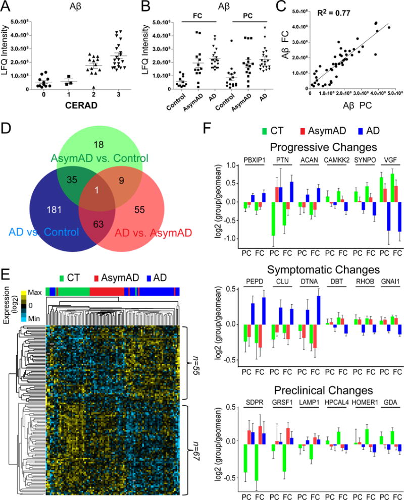
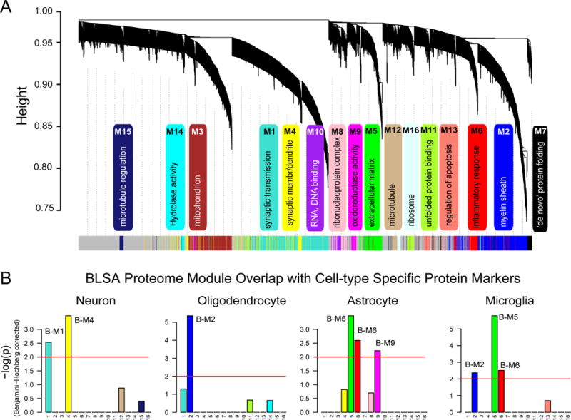
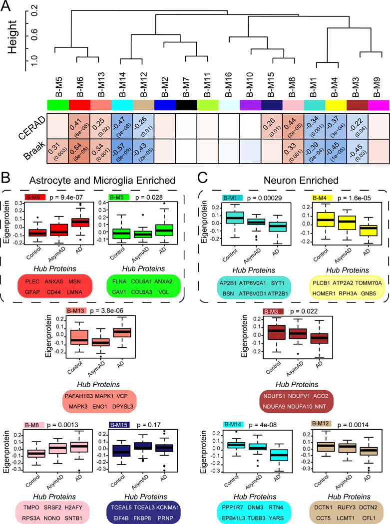
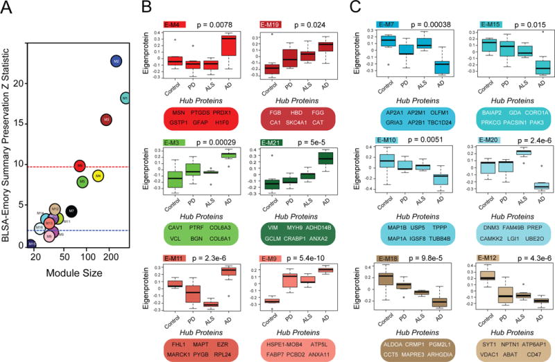
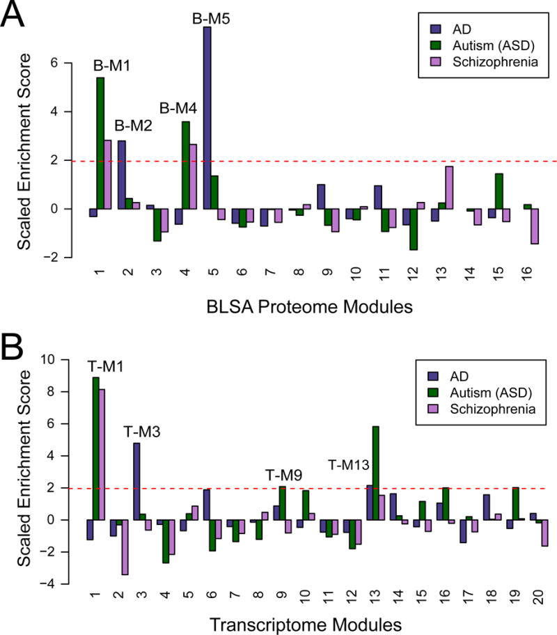
Here, we report proteomic analyses of 129 human cortical tissues to define changes associated with the asymptomatic and symptomatic stages of Alzheimer's disease (AD). Network analysis revealed 16 modules of co-expressed proteins, 10 of which correlated with AD phenotypes. A subset of modules overlapped with RNA co-expression networks, including those associated with neurons and astroglial cell types, showing altered expression in AD, even in the asymptomatic stages. Overlap of RNA and protein networks was otherwise modest, with many modules specific to the proteome, including those linked to microtubule function and inflammation. Proteomic modules were validated in an independent cohort, demonstrating some module expression changes unique to AD and several observed in other neurodegenerative diseases. AD genetic risk loci were concentrated in glial-related modules in the proteome and transcriptome, consistent with their causal role in AD. This multi-network analysis reveals protein- and disease-specific pathways involved in the etiology, initiation, and progression of AD.


Figures

Fig. 1
Fig. 1. Differential abundance of Aβ and other proteins observed in AsymAD and AD brain
(A and B) Aβ measurements measured by LFQ ion intensity correlate with CERAD and disease status in precuneus (PC) and frontal cortex (FC). (C) Correlation for Aβ levels across 47 paired samples in the PC and FC. (D) Venn diagram showing a total of 362 unique proteins in both brain regions (FC and PC) that were determined to be significantly altered (decreased or increased) by ANOVA followed by Tukey’s post-hoc test (p <0.01) in the three comparisons. (E) Supervised hierarchical clustering of 123 significant proteins altered in FC and PC by criteria described in methods. (F) Several significant proteins in AD displayed a progressive change across comparisons of control, AsymAD and AD groups (top panel), whereas other proteins were significantly changed only during the symptomatic phase of AD (middle panel). Protein markers which trended as changing in both the preclinical and symptomatic stage of AD (bottom panel) compared to control group(s).
Fig. 2
Fig. 2. Protein co-expression classifies the proteome into modules associated with specific gene ontologies and brain cell types
(A) WGCNA cluster dendrogram groups proteins (n=2,735) measured across FC and PC into distinct protein modules (M1–16) defined by dendrogram branch cutting. These modules were significantly enriched for gene ontologies linked to discrete cellular functions and/or organelles in the brain. (B) Cell type enrichment was assessed by cross-referencing module proteins (via matching gene symbols) using the one-tailed Fisher’s exact test against lists of proteins determined as enriched in neurons, oligodendrocytes, astrocytes and microglia (Table S4). The FDR was corrected for multiple comparisons by the Benjamini-Hochberg (BH) method, bars extending above the line represent BH p<0.01.
Fig. 3
Fig. 3. BLSA protein modules correlate to cognitive status and AD neuropathological burden
(A) BLSA protein modules were clustered to assess module relatedness based on correlation of protein co-expression eigenproteins. Pearson correlation and P value between module eigenprotein expression and CERAD (top) and Braak (bottom). (B) Module expression profiles and key hub proteins that are positively correlated with CERAD and/or Braak. (C) Module expression profiles and key hub proteins that are negatively correlated with CERAD and/or Braak. Box plots with error bars are displayed for each of the three groups of case samples (control, AsymAD and AD). Significance was measured using one-way nonparametric ANOVA, Kruskal-Wallis p-values. Cell type associated modules are encircled (dashed line).
Fig. 4
Fig. 4. BLSA network changes are preserved in the Emory proteome
(A) In the BLSA proteomic network, 13 of the 16 modules were highly preserved in the Emory proteomic network, with Zsummary scores above 2 (p<0.05). Larger modules were some of the most preserved (above red line, p<0.01). (B) Emory protein modules and top hub proteins that increased in AD selectively or across all neurodegenerative diseases. (C) Emory protein modules and top hub proteins that decreased in AD selectively or across all neurodegenerative diseases. Significance was measured using one-way nonparametric ANOVA, Kruskal-Wallis p-values.
Fig. 5
Fig. 5. Overlap between RNA and protein co-expression networks in AD
(A) A hypergeometric two-tailed Fisher’s exact test was used to determine which modules shared significant overlap or depletion of module members between the Emory and BLSA proteome networks (left panel) and Emory proteome and RNA networks (right panel). The 23 modules in the Emory case network (x-axis), clustered by module eigenprotein relatedness, were aligned to the 16 modules in the BLSA network (y-axis). Module gene symbol lists showed either significant overlap (red), depletion (blue) or no significant under- or over-representation (white) in protein membership. Numbers are positive signed −log10(FDR-corrected overrepresentation p values) representing degree of significance of overlap; asterisks also represent degree of significance for either positive or depleted comparisons: *, p<0.05; **, p<0.01, ***, p<0.005. (B) The degree of significance related to AD with correlation sign [signed −log10(FDR corrected p value)] is provided for each transcriptome module. Module eigengenes were correlated to AD status and multiple comparisons were accounted for by FDR correction (Benjamini-Hochberg) across modules with significance. (C) RNA modules were found enriched for specific cell type markers (Table S4) including neuronal, oligodendrocyte, astrocyte, microglia and endothelial cells following one-way Fisher’s exact test overlap with cell-type specific transcriptomes. (D) Pearson correlation analysis between all overlapping protein-RNA targets (n=2,406, left panel). Pearson correlation analysis between Protein-RNA targets in Emory modules that overlap with the transcriptome and change in the AD proteome (n=411, middle panel). This included protein modules increased in AD (E-M4, E-M21 and E-M3) that are enriched in astrocyte/microglia/endothelial markers and modules decreased in AD (E-M15 and E-M7) that are enriched with neuronal markers. Pearson correlation analysis between Protein-RNA targets in Emory modules that did not overlap with modules in the transcriptome (n=411, right panel), yet were increased (E-M19, E-M11 and E-M9) or decreased (E-M20, E-M10, E-M6, E-M18 and E-M12) in AD. Genes and cognate proteins were grouped and colored by their Emory protein module membership.
Fig. 6
Fig. 6. AD GWAS candidates are over-represented in protein and RNA networks associated glial cell types
(A) AD GWAS candidate genes (darkslateblue) in the BLSA network were found significantly over-represented in oligodendrocyte module (B-M2), and in the astrocyte/microglia (B-M5) module. GWAS risk candidate genes for both ASD (darkgreen) and schizophrenia (magenta) were significantly over-represented in the B-M1 and B-M4 neuron/synaptic modules (B) AD GWAS candidate genes in the transcriptome were overrepresented microglial/endothelial modules T-M3 and T-M18 and an oligodendrocyte associated module (T-M13). Similar to the proteome, ASD and schizophrenia GWAS targets were significantly over-represented in neuronal transcriptome modules (T-M1 and T-M9). ASD was also found in the oligodendrocyte enriched T-M13 module. Random sampling (10,000 times) of the MAGMA gene list was used to assess the significance of the module enrichment score (* p value <0.05).

Comment in

References

    1. Abreu RDS, Penalva LO, Marcotte EM, Vogel C. Global signatures of protein and mRNA expression levels. Molecular bioSystems. 2009;5:1512–1526. - PMC - PubMed
    1. Alexander GE, Chen K, Pietrini P, Rapoport SI, Reiman EM. Longitudinal PET Evaluation of Cerebral Metabolic Decline in Dementia: A Potential Outcome Measure in Alzheimer’s Disease Treatment Studies. American Journal of Psychiatry. 2002;159:738–745. - PubMed
    1. Bai B, Hales CM, Chen PC, Gozal Y, Dammer EB, Fritz JJ, Wang X, Xia Q, Duong DM, Street C, et al. Proceedings of the National Academy of Sciences. 2013. U1 small nuclear ribonucleoprotein complex and RNA splicing alterations in Alzheimer’s disease. - PMC - PubMed
    1. Braak H, Braak E. Neuropathological stageing of Alzheimer-related changes. Acta Neuropathol. 1991;82:239–259. - PubMed
    1. Cerami E, Demir E, Schultz N, Taylor BS, Sander C. Automated network analysis identifies core pathways in glioblastoma. PloS one. 2010;5:e8918. - PMC - PubMed

Publication types

MeSH terms