

Is There a Magnetic Resonance Imaging-Discernible Cause for Trigeminal Neuralgia? A Structured Review
- PMID: 27989975
- PMCID: PMC5326610
- DOI: 10.1016/j.wneu.2016.10.104
Is There a Magnetic Resonance Imaging-Discernible Cause for Trigeminal Neuralgia? A Structured Review
Abstract
Background: Trigeminal neuralgia (TN) is a chronic brain condition involving the trigeminal nerve and characterized by severe and recurrent facial pain. Although the cause of TN has been researched extensively, there is a lack of convergence on the physiologic processes leading to pain symptoms. This review seeks to better elucidate the underlying pathophysiology of TN by analyzing the outcomes of studies that use magnetic resonance structural imaging and diffusion-weighted imaging to examine nerve damage in patients with TN.
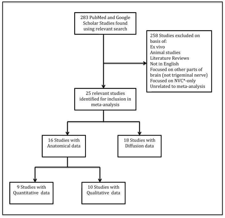
Methods: Performing a structured review of the literature, the authors included human magnetic resonance anatomic and diffusion-weighted imaging studies aimed at visualizing the trigeminal nerve or measuring neural damage pertaining to TN. Studies that measured and compared nerve damage in the affected and unaffected sides in patients or patients and controls were analyzed for neural changes associated with TN.
Results: Twenty-five studies met inclusion criteria. Overall, the data from the anatomic and diffusion studies showed decreased volume and cross-sectional area, decreased fractional anisotropy, and increased apparent diffusion coefficient and diffusivity associated with the affected side of patients compared with the unaffected side as well as in patients compared with controls.
Conclusions: A review of the studies included indicates that neural differences exist between the affected and unaffected sides in patients as well as between patients and controls in both structural and diffusion metrics. The amalgamated data suggest that damage of the trigeminal nerve tissue is commonly found in patients with TN and could be a primary factor in TN pathophysiology.
Keywords: Diffusion-weighted imaging; Magnetic resonance imaging; Nerve damage; Trigeminal neuralgia.
Copyright © 2016 Elsevier Inc. All rights reserved.
Conflict of interest statement
Conflicts of interest: none
Figures
Similar articles
-
7.0 Tesla MRI tractography in patients with trigeminal neuralgia.Magn Reson Imaging. 2018 Dec;54:265-270. doi: 10.1016/j.mri.2017.12.033. Epub 2018 Jan 3. Magn Reson Imaging. 2018. PMID: 29305127
-
Microstructural alterations in trigeminal neuralgia determined by diffusion tensor imaging are independent of symptom duration, severity, and type of neurovascular conflict.J Neurosurg. 2016 Mar;124(3):823-30. doi: 10.3171/2015.2.JNS142587. Epub 2015 Sep 25. J Neurosurg. 2016. PMID: 26406792
-
Trigeminal neuralgia due to neurovascular compression: high-spatial-resolution diffusion-tensor imaging reveals microstructural neural changes.Radiology. 2011 Feb;258(2):524-30. doi: 10.1148/radiol.10100477. Epub 2010 Nov 9. Radiology. 2011. PMID: 21062923
-
Recent Advances of Magnetic Resonance Neuroimaging in Trigeminal Neuralgia.Curr Pain Headache Rep. 2021 Apr 6;25(6):37. doi: 10.1007/s11916-021-00957-0. Curr Pain Headache Rep. 2021. PMID: 33821366 Review.
-
Advanced neuroimaging of the trigeminal nerve and the whole brain in trigeminal neuralgia: a systematic review.Pain. 2025 Feb 1;166(2):282-310. doi: 10.1097/j.pain.0000000000003365. Epub 2024 Aug 8. Pain. 2025. PMID: 39132931
Cited by
-
A combined radiomics and anatomical features model enhances MRI-based recognition of symptomatic nerves in primary trigeminal neuralgia.Front Neurosci. 2024 Oct 24;18:1500584. doi: 10.3389/fnins.2024.1500584. eCollection 2024. Front Neurosci. 2024. PMID: 39513045 Free PMC article.
-
The trigeminal pathways.J Neurol. 2022 Jul;269(7):3443-3460. doi: 10.1007/s00415-022-11002-4. Epub 2022 Mar 6. J Neurol. 2022. PMID: 35249132 Review.
-
The Challenges in Clinical Diagnosis of Trigeminal Neuralgia: A Review.Cureus. 2024 Jun 7;16(6):e61898. doi: 10.7759/cureus.61898. eCollection 2024 Jun. Cureus. 2024. PMID: 38978896 Free PMC article. Review.
-
Low Field MRI Measurements of the Normal Canine Trigeminal Nerve.Front Vet Sci. 2020 May 19;7:274. doi: 10.3389/fvets.2020.00274. eCollection 2020. Front Vet Sci. 2020. PMID: 32509809 Free PMC article.
-
Conservative Management of Trigeminal Neuralgia and Degenerative Cervical Myelopathy: A Case Report.Cureus. 2024 Feb 29;16(2):e55274. doi: 10.7759/cureus.55274. eCollection 2024 Feb. Cureus. 2024. PMID: 38558660 Free PMC article.
References
-
- Pollock BE, Ecker RD. A prospective cost-effectiveness study of trigeminal neuralgia surgery. Clin J Pain. 2005;21(4):317–22. - PubMed
-
- Harsha KJ, et al. Imaging of vascular causes of trigeminal neuralgia. J Neuroradiol. 2012;39(5):281–9. - PubMed
-
- Katusic S, et al. Incidence and clinical features of trigeminal neuralgia, Rochester, Minnesota, 1945–1984. Annals of neurology. 1990;27(1):89–95. - PubMed
-
- Pamir MN, et al. Microvascular decompression in the surgical management of trigeminal neuralgia. Neurosurg Rev. 1995;18(3):163–7. - PubMed
Publication types
MeSH terms
Grants and funding
LinkOut - more resources
Full Text Sources
Other Literature Sources
Medical
Miscellaneous
