Review
. 2017 Feb;13(2):115-131.
doi: 10.1038/nrneph.2016.182. Epub 2016 Dec 19.

The renal Fanconi syndrome in cystinosis: pathogenic insights and therapeutic perspectives

Affiliations
Review

The renal Fanconi syndrome in cystinosis: pathogenic insights and therapeutic perspectives

Stephanie Cherqui et al. Nat Rev Nephrol. 2017 Feb.

Abstract

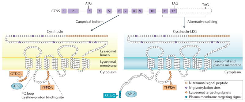
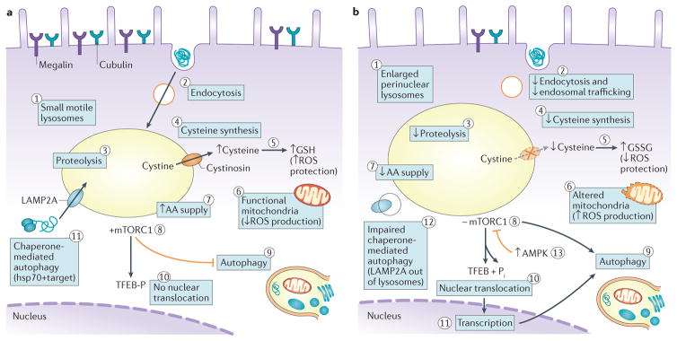
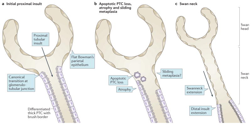
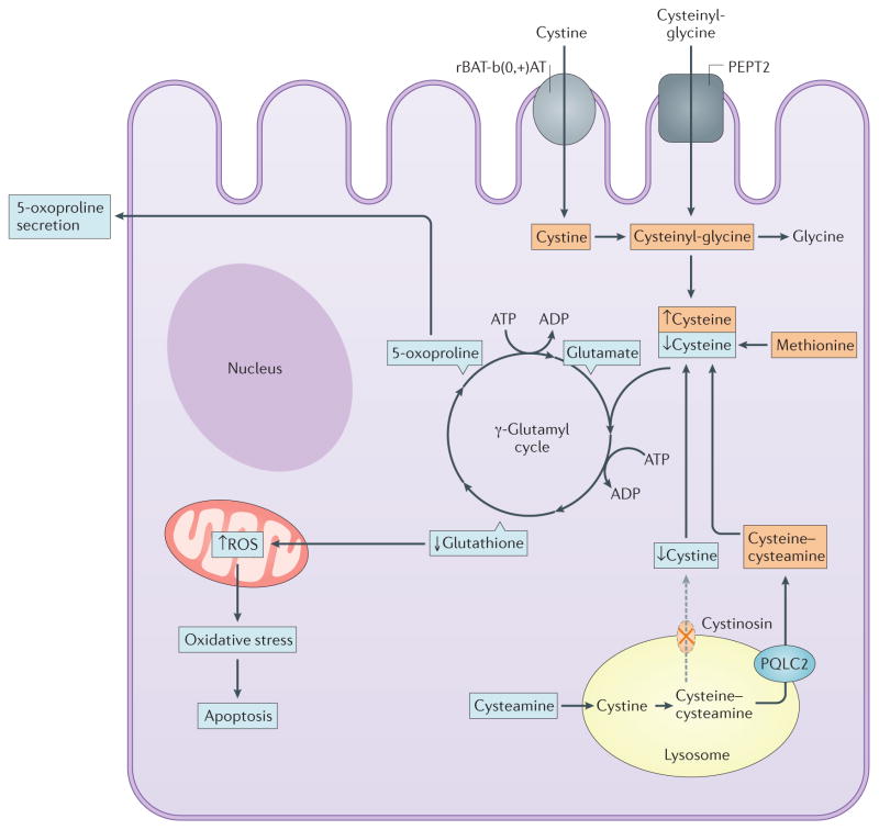
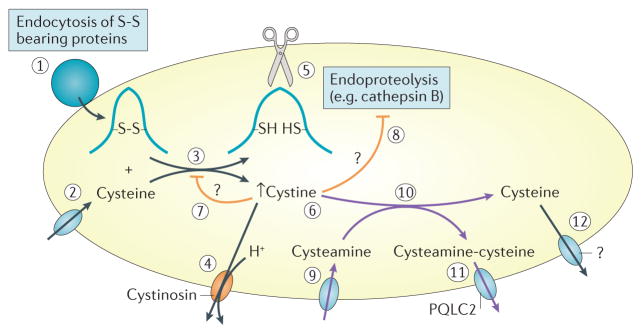
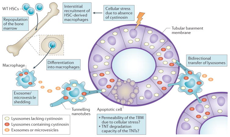
Cystinosis is an autosomal recessive metabolic disease that belongs to the family of lysosomal storage disorders. It is caused by a defect in the lysosomal cystine transporter, cystinosin, which results in an accumulation of cystine in all organs. Despite the ubiquitous expression of cystinosin, a renal Fanconi syndrome is often the first manifestation of cystinosis, usually presenting within the first year of life and characterized by the early and severe dysfunction of proximal tubule cells, highlighting the unique vulnerability of this cell type. The current therapy for cystinosis, cysteamine, facilitates lysosomal cystine clearance and greatly delays progression to kidney failure but is unable to correct the Fanconi syndrome. This Review summarizes decades of studies that have fostered a better understanding of the pathogenesis of the renal Fanconi syndrome associated with cystinosis. These studies have unraveled some of the early molecular changes that occur before the onset of tubular atrophy and identified a role for cystinosin beyond cystine transport, in endolysosomal trafficking and proteolysis, lysosomal clearance, autophagy and the regulation of energy balance. These studies have also led to the identification of new potential therapeutic targets and here, we outline the potential role of stem cell therapy for cystinosis and provide insights into the mechanism of haematopoietic stem cell-mediated kidney protection.


Conflict of interest statement

Competing interests

The authors declare no competing interests.

Figures

Figure 1
Figure 1. CTNS produces two isoforms with distinct subcellular localizations
CTNS, which encodes the seven transmembrane lysosomal protein cystinosin, is composed of 12 exons (solid boxes) with the start codon (ATG) in exon 3 and two alternative stop codons (TAGs) in exon 12. The common, canonical form of cystinosin is shown on the left. The 3’ region of CTNS (dashed box) encodes the C-terminal extension of the less frequent isoform, cystinosin-LKG, which arises from alternative splicing in exon 12 (right). Canonical cystinosin exclusively localizes to lysosomes due to the concerted effect of two lysosome-targeting signals: a classic tyrosine-based C-terminal motif, GYDQL, and a non-classic motif, YFPQA, in the fifth transmembrane loop named the ‘PQ loop’, which also contains a cystine proton binding site. The GYDQL motif allows recognition by the adaptor protein complex 3 (AP 3), which is responsible for direct lysosomal targeting of cystinosin. The cystinosin-LKG isoform lacks the GYDQL motif but instead contains the carboxyl-terminal SSLKG sequence, which directs this isoform to the plasma membrane, with secondary endocytic trafficking to lysosomes involving AP-2 complexes.
Figure 2
Figure 2. Cystine crystals accumulate within lysosomes of cystinotic cells
a | Conventional electron micrograph of a proximal tubule cell from an 8-month old Ctns-knockout mouse showing a lysosome that is about twofold larger in diameter than usual. b | Pseudo-colour conversion of grey levels, to better demonstrate intertwining of the normal dense matrix (dark areas) with amorphous, less electron-dense material suggestive of undigested protein (blue areas); and the two types of crystals: a sharp, needle-shaped crystal (arrow) and two polyhedric crystals (arrow heads). Such crystals are never seen before 6 months of age and needles do not usually contact the lysosomal membrane. Note the lack of luminal membrane packing that identifies autophagic structures. c | Conventional electron micrograph of a mouse proximal tubule cell deformed by a single huge lysosome. Red lines indicate the straight borders and angles of a polyhedric crystal, close to 10 μm in height. Note the preservation of the brush border in adjacent cells. Part c reproduced with permission from the American Society of Nephrology © Gaide Chevronnay, H. P. et al. J. Am. Soc. Nephrol. 25, 1256–1269 (2014).
Figure 3
Figure 3. Cell biological alterations in cystinotic cells
a | Lysosomes of healthy proximal tubule cells (PTCs) are small, motile and dispersed throughout the cytoplasm (1), where they can readily fuse with late endosomes. High apical expression of the tandem multiligand receptors, megalin and cubilin, and their unusually fast recycling rate result in extremely active receptor mediated endocytosis (2). Endosomal trafficking, transfer into lysosomes and proteolysis (3) of ultrafiltrated disulfide rich plasma proteins ensures an ample lysosomal supply of amino acids (AAs) together with sulfur-bearing cystine. Following cystinosin-mediated export from the lysosome, cystine becomes a source of cysteine in the cystosol (4), which favours the biosynthesis of reduced glutathione (GSH), which confers protection against reactive oxygen species (ROS; 5). Moreover, functional mitochondria do not release ROS (6). An abundance of AAs (7) keeps the mTORC1 complex at the lysosomal membrane in its active kinase form (8). Crucial physical interactions between cystinosin and mTORC1 are not indicated. Lysosomal mTORC1 inhibits macroautophagy (9) and maintains TFEB phosphorylation, thereby abolishing its nuclear translocation and transcriptional effects (10). Localization of LAMP2A at the lysosomal membrane supports the clearance of altered cytosolic proteins by chaperone-mediated autophagy (11). b | In cystinotic PTCs, lysosomes engorged by undigested proteins (evident as amorphous inclusions) become enlarged and cluster around nuclei (1); they are less motile than lysosomes of healthy PTCs and are located further from the apical membrane, rendering them less accessible to endocytic cargo and less prone to exocytic clearance. Endocytic uptake is also decreased as expression of megalin and cubilin declines (2). Impaired proteolysis (3) lowers AA supply (7). Moreover, cystine can no longer cross the lysosomal membrane due to an absence or loss-of-function of cystinosin, which presumably limits cysteine availability (4) and therefore GSH synthesis, reversing the ratio of GSH to oxidized glutathione (GSSG), leading to increased oxidative stress by ROS (5). Concomitantly, ROS production increases as mitochondria are damaged and/or are less efficiently cleared (6). The shortage of free AAs (7) inhibits mTORC1 (8), which activates autophagic programmes, including mitophagy (9). mTORC1 inactivation also leads to dephosphorylation and nuclear translocation of TFEB (10), activating transcription programmes (11) that further promote autophagy. Chaperone-mediated autophagy of altered (including oxidized) cytosolic proteins is hampered, in part because of impaired LAMP2A trafficking to lysosomes (12). Continuous autophagy eventually causes cell atrophy. In parallel, energy depletion activates AMPK, which inhibits mTORC1 thus activates autophagy (13). Enhanced apoptosis is not represented. Hsp70, heat shock protein-70.
Figure 4
Figure 4. Origin and consequences of swan neck deformities
a | The canonical localization at the glomerulotubular junction represents the transition between the flat parietal epithelium typical of the Bowman capsule and the thick columnar epithelium lining the proximal tubules, with a highly differentiated apical pole represented by a brush border. The transition between the two types of epithelial cells can occur within the glomerulus, with the lower third of the Bowman capsule lined by thick epithelium that is undistinguishable from differentiated proximal tubule cells (PTCs). The opposite scenario (that is, a transition from thin to thick epithelium distal to the glomerulotubular junction) is never found under normal conditions. The insult here arises from the apical pole by an ultrafiltrated and reabsorbed toxic factor. We suggest that this factor could be disulfide-bearing albumin, which is the main source of cystine, and first affects the most proximal PTCs. b | Swan neck deformities arise from the apoptotic cell death and shedding of individual PTCs; atrophy by dedifferentiation of PTCs; and possibly by replacement of dead PTCs upon expansion of the flat Bowman epithelium from the glomerular wall into the tubules (sliding metaplasia hypothesis). c | The characteristic features of a microdissected nephron exhibiting a normal-sized glomerulus (swan head) and an atrophic proximal tubule (swan neck) suggest the extension of lesions to cells at the base of the neck, as they become exposed to the ultrafiltrated and reabsorbed toxic factor, are thus governed by the same mechanisms as in part b.
Figure 5
Figure 5. Cystinosin deficiency and oxidative stress in proximal tubular cells
Absence of cystinosin was thought to lead to decreased cytosolic cysteine content, resulting in decreased glutathione and increased production of reactive oxygen species (ROS) and oxidative stress (blue squares). One of intermediates of the γ-glutamyl cycle is 5-oxoproline, which is increased in the urine of cystinotic patients. However, alternative sources of cysteine exist including through the reabsorption of cysteinyl-glycine by PEPT2 and cystine by the actions of the amino-acid transporter rBAT-b(0,+)AT (orange squares). Moreover, cysteamine drug, another source of cytoplasmic cysteine, does not prevent the renal Fanconi syndrome, suggesting the contribution of factors other than cysteine deficiency to this phenotype.
Figure 6
Figure 6. Lysosomal membrane transport and luminal events associated with cystinosis and cysteamine therapy
Under normal conditions (black pathways (left)), vesicular transfer into lysosomes of disulfide-bearing proteins, such as albumin (1), which is avidly taken up by receptor-mediated endocytosis into proximal tubule cells, combined with lysosomal import of cysteine by a still unknown mechanism (2) leads to the reduction of disulfide bonds, resulting in protein unfolding and release of free cystine (3),. Cystine is rapidly exported into the cytosol by the cystine proton symporter, cystinosin (4). Protein unfolding unmasks peptide bonds buried in globular proteins thus allows endoproteolytic attack by the concerted action of lysosomal cathepsins, such as the cysteine protease, cathepsin B (5). Free thiol in the catalytic site of cysteine proteinases is preserved by the reducing lysosomal environment. In the absence of cystinosin (orange pathways), cystine export across the lysosomal membrane is abolished. Cystine accumulation (6) inhibits the reduction of disulfide bonds in lysosomes, which thereby is expected to slow down further cystine generation (7) but also impairs protein unfolding and inactivates cysteine proteases (8), together leading to impairment of proteolysis (8). Cysteamine (β-mercaptoethylamine; purple pathways (right)) penetrates lysosomes by a distinct importer (9) and reacts with cystine to exchange its disulfide bridge into a mixed (cysteamine cysteine) disulfide (10), which exits via the cationic amino-acid exporter PQLC2 (11), and generates free cysteine, which must also exit lysosomes although this transporter is unknown (12). Question marks indicate that steps 7, 8 and 12 are still hypothetical.
Figure 7
Figure 7. Proposed mechanisms by which haematopoietic stem cell (HSC) transplantation rescues cystinosis-induced Fanconi syndrome
Injury to renal cells, including proximal tubular cells (PTCs) induced by loss of cystinosin triggers an inflammatory response, which probably aggravates the injury. Transplanted wild type (WT) cystinosin sufficient HSCs repopulate the bone marrow of Ctns knockout mice then migrate into injured tissues, where they differentiate into macrophages. Extracellular exosome or microvesicle-mediated transfer of cystinosin might correct the cystinosin deficiency of adjacent interstitial renal cells. However, rescue of PTCs, which are embedded in the tubular basement membrane, requires tunnelling nanotubes (TNTs), which extend from the macrophages across the tubular basement membrane, and are capable of bidirectional transfer of lysosomes, cystinosin-containing lysosomes (red vesicles) from wild-type HSC-derived macrophages into PTCs and cystine-loaded lysosomes (white vesicles) from Ctns deficient PTCs into macrophages. A single macrophage can generate multiple TNTs that have the ability to transfer lysosomes over long distances, which makes this route of cross-correction particularly efficient, likely accounting for the observed long-term correction of the Fanconi syndrome in Ctns-knockout mice.

Similar articles

Cited by

References

    1. Fanconi G. Die nicht diabetischen glykosurien und hyperglykamiendes altem kindes. Jahrbuch Kinderheilkunde. 1931;133:257–300.
    1. De Toni G. Remarks on the relations between renal rickets (renal dwarfism) and renal diabetes. Acta Paediatr. 1933;16:479–484.
    1. Debre R, Marie J, Cleret J, Messimy R. Rachitisme tardif coexistant avec une nephrite chronique et une glycosurie. Arch Med Enfants. 1934;37:597–606.
    1. Haffner D, et al. Long-term outcome of paediatric patients with hereditary tubular disorders. Nephron. 1999;83:250–260. - PubMed
    1. Gahl WA, Thoene JG, Schneider JA. Cystinosis. N Engl J Med. 2002;347:111–121. - PubMed