. 2016 Dec 1:7:557.
doi: 10.3389/fimmu.2016.00557. eCollection 2016.

Oxidative Burst-Dependent NETosis Is Implicated in the Resolution of Necrosis-Associated Sterile Inflammation

Affiliations

Oxidative Burst-Dependent NETosis Is Implicated in the Resolution of Necrosis-Associated Sterile Inflammation

Mona H C Biermann et al. Front Immunol. .

Erratum in

Abstract

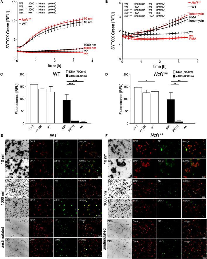
Necrosis is associated with a profound inflammatory response. The regulation of necrosis-associated inflammation, particularly the mechanisms responsible for resolution of inflammation is incompletely characterized. Nanoparticles are known to induce plasma membrane damage and necrosis followed by sterile inflammation. We observed that injection of metabolically inert nanodiamonds resulted in paw edema in WT and Ncf1** mice. However, while inflammation quickly resolved in WT mice, it persisted over several weeks in Ncf1** mice indicating failure of resolution of inflammation. Mechanistically, NOX2-dependent reactive oxygen species (ROS) production and formation of neutrophil extracellular traps were essential for the resolution of necrosis-induced inflammation: hence, by evaluating the fate of the particles at the site of inflammation, we observed that Ncf1** mice deficient in NADPH-dependent ROS failed to generate granulation tissue therefore being unable to trap the nanodiamonds. These data suggest that NOX2-dependent NETosis is crucial for preventing the chronification of the inflammatory response to tissue necrosis by forming NETosis-dependent barriers between the necrotic and healthy surrounding tissue.

Keywords: NETosis; inflammation; nanodiamonds; necrosis; reactive oxygen species; resolution.


Conflict of interest statement

The authors declare that the research was conducted in the absence of any commercial or financial relationships that could be construed as a potential conflict of interest.

Figures

Figure 1
Figure 1
Diamond nanoparticles induce plasma membrane damage and NET formation in human leukocytes. (A) Assessment of DNA exposure in neutrophils (PMN) and peripheral blood mononuclear cells (PBMC). Cells without stimulus (wo) or incubated with nanodiamonds (10 nm) or microdiamonds (1000 nm) were measured using SYTOX Green. (B) Exposure of DNA by neutrophils was measured in response to phorbol 12-myristate 13-acetate (PMA) to nanodiamonds (10 nm) or microdiamonds (1000 nm) and without stimulus (wo) using SYTOX Green. (C) Dose-dependent increase of DNA exposure in PMN without stimulus (wo) or incubated with nanodiamonds (10 nm) or microdiamonds (1000 nm) by SYTOX Green after 150 min. (D) Microscopic analysis of PMN incubated with nanodiamonds (10 nm) or microdiamonds (1000 nm) and stained for DNA (Hoechst33342), neutrophil elastase (NE), or citrullinated histone H3 (citH3). Diamonds are visible in differential interference contrast (DIC) images. Cy5 fluorescence was artificially colored green. (E) Microscopic analysis of PBMC incubated with nanodiamonds (10 nm) or microdiamonds (1000 nm) and stained for DNA (Hoechst33342) and CD45-FITC. Diamonds are visible in DIC and provide strong background fluorescence in FITC. (F) Quantification of extracellular DNA and citH3 in human PMN without stimulus (wo) or incubated with nanodiamonds (10 nm) or microdiamonds (1000 nm) after 240 min. Significances below 0.001 are depicted. (G) Microscopic analysis of PMN incubated with nanodiamonds and treated with the ROS scavenger N-acetyl cysteine stained for DNA (Hoechst33342), NE, or citH3. Diamonds are visible in DIC. (H) In silico quantification of microscopic pictures regarding the number and area of DNA (Hoechst33342)-stained NET structures (AggNET) formed by PMN incubated with nanodiamonds in the absence or presence of the ROS scavenger N-acetyl cysteine. Each dot represents one analyzed field of view (FOV). (I) Microscopic analysis of NET formation in response to nanodiamonds induced necrosis of CFSE-labeled PBMC stained for DNA (Hoechst33342), NE, or citH3. Diamonds are visible in DIC. Data of one representative experiment reflecting the result of three independent experiments are shown as medians with interquartile ranges of triplicates. Two-way ANOVA (A–C), one-way ANOVA (F), Kruskal–Wallis one-way analysis of variance (H), and Mann–Whitney U test (I) were used to evaluate differences among means; **p < 0.05, ***p < 0.001, and relative fluorescence units (RFU) field of view (FOV).
Figure 2
Figure 2
Diamond nanoparticles induce plasma membrane damage and NET formation in murine bone marrow cells. (A) Analysis of DNA exposure by bone marrow cells of WT and Ncf1** mice in response to nanodiamonds (10 nm) or microdiamonds (1000 nm) or without stimulus (wo) using SYTOX Green. (B) Analysis of DNA exposure by bone marrow cells of WT and Ncf1** mice in response to NET-inducing stimuli (PMA and ionomycin) or without stimulus (wo) using SYTOX Green. (C) Quantification of extracellular DNA and citrullinated histone H3 (citH3) in bone marrow cells of WT mice without stimulus (wo) or incubated with nanodiamonds (10 nm) or microdiamonds (1000 nm) after 240 min. (D) Quantification of extracellular DNA and citH3 in bone marrow cells of Ncf1** mice without stimulus (wo) or incubated with nanodiamonds (10 nm) or microdiamonds (1000 nm) after 240 min. (E) Microscopic analysis of bone marrow cells of WT mice incubated with nanodiamonds (10 nm) or microdiamonds (1000 nm) and unstimulated cells stained for DNA (Hoechst33342), neutrophil elastase (NE), or citH3. Diamonds are visible in differential interference contrast (DIC). Cy5 fluorescence was artificially colored green. (F) Microscopic analysis of bone marrow cells of Ncf1** mice incubated with of nanodiamonds (10 nm) or microdiamonds (1000 nm) and unstimulated cells stained for DNA (Hoechst33342), NE, or citH3. Diamonds are visible in DIC. Cy5 fluorescence was artificially colored green. Data of one representative experiment reflecting the result of three independent experiments are shown as medians with interquartile ranges of triplicates. Two-way ANOVA (A,B) and one-way ANOVA (C,D) were used to evaluate differences among means; *p < 0.05, **p < 0.01, ***p < 0.001, and relative fluorescence units (RFU).
Figure 3
Figure 3
Chronification of necrosis-associated sterile inflammation in NOX2-deficient mice. (A) Paw edema in response to injected nanodiamonds in WT and Ncf1** mice. Means ± SEM of the relative hind paw thickness are shown. #p < 0.05 of areas under the curves as determined by t test; n = 4–6. (B) Measurement of hind paw edema in response to injected microdiamonds in WT and Ncf1** mice. Means ± SEM of the relative hind paw thickness are shown. (C) Measurement of hind paw edema in response to injected nanodiamonds in WT mice in the absence or presence of DNase I. Means ± SEM of the relative hind paw thickness are shown. *p < 0.001; n = 5 (D) Paws of WT and Ncf1** mice microdissected 15 days after injection of nanodiamonds (dark gray matter, 10 nm) and of microdiamonds (white matter, 1000 nm). (E) Macroscopic pictures of dissected paws of WT and Ncf1** mice at day 28 after injection of nanodiamonds showing nanodiamonds (dark gray matter) on the paw and overlaying skin. (F) Paraffin sections of overlaying skins shown in (E), stained for neutrophil elastase (NE), citrullinated histone H3 (citH3), and DNA (PI) as well as differential interference contrast (DIC). Nanodiamonds giving highest contrast in DIC were artificially colored blue. Data are shown as means ± SEM. Two-way ANOVA with Bonferroni post testing (A–C) was used to evaluate differences among means.
Figure 3
Figure 3
Chronification of necrosis-associated sterile inflammation in NOX2-deficient mice. (A) Paw edema in response to injected nanodiamonds in WT and Ncf1** mice. Means ± SEM of the relative hind paw thickness are shown. #p < 0.05 of areas under the curves as determined by t test; n = 4–6. (B) Measurement of hind paw edema in response to injected microdiamonds in WT and Ncf1** mice. Means ± SEM of the relative hind paw thickness are shown. (C) Measurement of hind paw edema in response to injected nanodiamonds in WT mice in the absence or presence of DNase I. Means ± SEM of the relative hind paw thickness are shown. *p < 0.001; n = 5 (D) Paws of WT and Ncf1** mice microdissected 15 days after injection of nanodiamonds (dark gray matter, 10 nm) and of microdiamonds (white matter, 1000 nm). (E) Macroscopic pictures of dissected paws of WT and Ncf1** mice at day 28 after injection of nanodiamonds showing nanodiamonds (dark gray matter) on the paw and overlaying skin. (F) Paraffin sections of overlaying skins shown in (E), stained for neutrophil elastase (NE), citrullinated histone H3 (citH3), and DNA (PI) as well as differential interference contrast (DIC). Nanodiamonds giving highest contrast in DIC were artificially colored blue. Data are shown as means ± SEM. Two-way ANOVA with Bonferroni post testing (A–C) was used to evaluate differences among means.
Figure 4
Figure 4
Neutrophils shield necrotic tissue by the formation of NETs building an anti-inflammatory barrier. Nanodiamonds induce necrosis by cell membrane damage, release of damage-associated molecular patterns, and the recruitment of neutrophils. Concerted NETosis of neutrophils then builds a barrier around the necrotic core consisting of aggregated NETs, which secludes the nanodiamonds and allows resolution of inflammation.

Similar articles

Cited by

References

    1. Kono H, Rock KL. How dying cells alert the immune system to danger. Nat rev Immunol (2008) 8(4):279–89.10.1038/nri2215 - DOI - PMC - PubMed
    1. Matzinger P. The danger model: a renewed sense of self. Science (2002) 296(5566):301–5.10.1126/science.1071059 - DOI - PubMed
    1. Scaffidi P, Misteli T, Bianchi ME. Release of chromatin protein HMGB1 by necrotic cells triggers inflammation. Nature (2002) 418(6894):191–5.10.1038/nature00858 - DOI - PubMed
    1. Huebener P, Pradere JP, Hernandez C, Gwak GY, Caviglia JM, Mu X, et al. The HMGB1/RAGE axis triggers neutrophil-mediated injury amplification following necrosis. J Clin Invest (2015) 125(2):539–50.10.1172/JCI76887 - DOI - PMC - PubMed
    1. Witko-Sarsat V, Rieu P, Descamps-Latscha B, Lesavre P, Halbwachs-Mecarelli L. Neutrophils: molecules, functions and pathophysiological aspects. Lab Invest (2000) 80(5):617–53.10.1038/labinvest.3780067 - DOI - PubMed