. 2016:2016:3094783.
doi: 10.1155/2016/3094783. Epub 2016 Nov 20.

Changes in IL-2 and IL-10 during Chronic Administration of Isoniazid, Nevirapine, and Paracetamol in Rats

Affiliations

Changes in IL-2 and IL-10 during Chronic Administration of Isoniazid, Nevirapine, and Paracetamol in Rats

Zanelle Bekker et al. Adv Pharmacol Sci. 2016.

Abstract

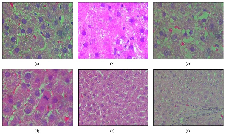
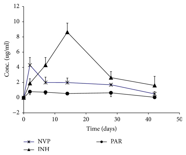
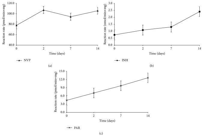
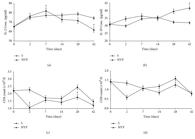
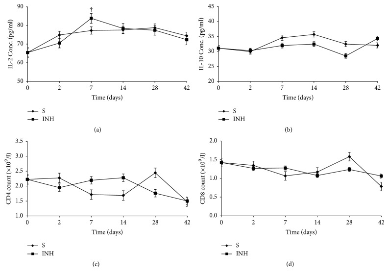
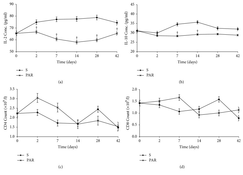
The aim of this study was to illustrate the initial subclinical drug-induced liver injury and the associated adaptive immune response by monitoring for the changes in plasma IL-2, IL-10, and some cytochrome P450 activity during chronic administration of nevirapine (NVP), isoniazid (INH), and paracetamol (PAR) in rats without clinical hepatotoxicity. Male Sprague-Dawley (SD) rats were divided into four groups (saline (S), NVP, INH, and PAR) of 25 animals each. The drugs were administered daily for 42 days at therapeutic doses (NVP 200 mg/kg, PAR 500 mg/kg, and INH 20 mg/kg) to the respective groups by oral gavage and five rats per group were sacrificed weekly. All the three drugs induced a subclinical liver injury in the first 2-3 weeks followed by healing, indicating adaption. The liver injury was pathologically similar and was associated with immune stimulation and increased cytochrome P450 activity. NVP- and PAR-induced liver injury lasted up to 14 days while that for INH lasted for 28 days. NVP-induced liver injury was associated with increased IL-2, CD4 count, and CYP3A2 activity, followed by increased IL-10 during the healing phase. In conclusion, the initial drug-induced subclinical liver injury, its spontaneous healing, and the associated adaptive immune response have been demonstrated.


Conflict of interest statement

The authors confirm that the funding did not lead to any conflict of interests regarding the publication of this manuscript and that there are no other possible conflicts of interests in the manuscript.

Figures

Figure 1
Figure 1
Representative histopathology slides (×20) of rat livers from control (untreated) and NVP treated rats and their respective pathology reports. (a) (No NVP): untreated group. “No pathology appears to be present in the control (untreated) animals”; (b) (NVP × 2 days): treated with NVP for 2 days. “Mild granular vacuolar degeneration and cell swelling, scattered cytonecrosis, nuclear pyknosis. Zonal necrosis... minimally present.”. (c) (NVP × 7 days): treated with NVP for 7 days. “vacuolar degeneration and cell swelling were moderate, single cell necrosis (cytonecrosis)... minimal centrilobular necrosis.”. (d) (NVP × 14 days): treated with NVP for 14 days. “moderate cellular swelling, vacuolar hepatopathy (degeneration) and granular cytoplasm... Hepatic parenchymal cell necrosis (cytonecrosis), and centrilobular zonal necrosis”. (e) (NVP × 28 days): treated with NVP for 28 days. “Granular vacuolar degeneration and cell swelling were minimal, as well as cytonecrosis.”. (f) (NVP × 42 days): treated with NVP for 42 days. “Centrilobular hepatocytes with minimal (1+) degeneration visible”.
Figure 2
Figure 2
Representative histopathology slides (×20) of rat livers from control (untreated) and isoniazid (INH) treated rats and their respective pathology reports. (a) (No INH): untreated group. “No pathology appears to be present in the control (untreated) animals”; (b) (INH × 2 days): treated with INH for 2 days. “Moderate granular vacuolar degeneration, loss of coordinated and well-organized hepatocytic cords. The cytoplasm appeared to be cloudy and granular, centrilobular zonal necrosis was minimal”. (c) (INH × 7 days): treated with INH for 7 days. “Severe degeneration of vacuoles and osmotic swelling of the mitochondria, cytonecrosis was moderate, loss of cell boundaries and nuclei”. (d) (INH × 14 days): treated with INH for 14 days. “Degenerative changes, cytonecrosis was present, disruption of the cytoplasm, minimal zonal necrosis was observed”. (e) (INH × 28 days): treated with INH for 28 days. “moderate cell swelling, mild cytonecrosis, hepatocytes appear swollen with granular cytoplasm.”. (f) (INH × 42 days): treated with INH for 42 days. “The histopathological lesions had improved, minimal cytonecrosis, centrilobular zonal necrosis was completely absent... one mitotic figure”.
Figure 3
Figure 3
Representative histopathology slides (×20) of rat livers from control (untreated) and PAR treated rats and their respective pathology reports. (a) (No PAR): section of rat liver from the untreated group: “No pathology appears to be present in the control (untreated) animals”; (b) (PAR × 2 days): PAR for 2 days. “moderate granular vacuolar degeneration and cell swelling, and minimal cytonecrosis, loss of cell boundaries, minimal mitosis, and minimal centrilobular zonal necrosis”. (c) (PAR × 7 days): PAR for 7 days: “mild degeneration and minimal cytonecrosis”. (d) (PAR × 14 days): PAR for 14 days. “moderate granular vacuolar degeneration and cell swelling, and minimal cytonecrosis loss of cell boundaries, minimal mitosis, and minimal hepatocyte mitosis”. (e) (PAR × 28 days): PAR for 28 days: “Hepatic cords with minimal vacuolar degenerative changes (×20)”; (f) (PAR × 42 days): PAR for 42 days: “Periportal hepatocytes, minimal degeneration visible (×20)”.
Figure 4
Figure 4
Plasma concentrations (mean ± sd) of NVP (stars), INH (triangles), and PAR (circles) in rats treated daily with NVP, INH, and PAR, respectively.
Figure 5
Figure 5
CYP3A2 activity in rats treated daily with NVP (a), CYP2E1 activity in rats treated daily with INH (b), and CYP1A2 activity in rats treated daily with PAR (c).
Figure 6
Figure 6
Serum concentrations (mean ± sd) of IL-2 (a), IL-10 (b), and CD4 (c) and CD8 (d) count during treatment with nevirapine (NVP) and saline (S). Note: † = p ≤ 0.05.
Figure 7
Figure 7
Serum concentrations (mean ± sd) of IL-2 (a), IL-10 (b), and CD4 (c) and CD8 (d) count during treatment with isoniazid (INH) and saline (S). Note: † = p ≤ 0.05.
Figure 8
Figure 8
Serum concentrations (mean ± sd) of IL-2 (a), IL-10 (b), and CD4 (c) and CD8 (d) count during treatment with paracetamol (PAR) and saline (S). Note: † = p ≤ 0.05.

Similar articles

Cited by

References

    1. Holt M. P., Ju C. Mechanisms of drug-induced liver injury. AAPS Journal. 2006;8(1):E48–E54. doi: 10.1208/aapsj080106. - DOI - PMC - PubMed
    1. Jaeschke H., Gores G. J., Cederbaum A. I., Hinson J. A., Pessayre D., Lemasters J. J. Mechanisms of hepatotoxicity. Toxicological Sciences. 2002;65(2):166–176. doi: 10.1093/toxsci/65.2.166. - DOI - PubMed
    1. Adams D. H., Ju C., Ramaiah S. K., Uetrecht J., Jaeschke H. Mechanisms of immune-mediated liver injury. Toxicological Sciences. 2010;115(2):307–321. doi: 10.1093/toxsci/kfq009. - DOI - PMC - PubMed
    1. Snodgrass W. R., Potter W. Z., Timbrell D. J., Mitchell J. R. Possible mechanisms of isoniazid-related hepatic injury. Clinical Research. 1975;22, article 323A
    1. Park B. K., Kitteringham N. R., Maggs J. L., Pirmohamed M., Williams D. P. The role of metabolic activation in drug-induced hepatotoxicity. Annual Review of Pharmacology and Toxicology. 2005;45:177–202. doi: 10.1146/annurev.pharmtox.45.120403.100058. - DOI - PubMed

LinkOut - more resources