

Agreement between Gonioscopic Examination and Swept Source Fourier Domain Anterior Segment Optical Coherence Tomography Imaging
- PMID: 27990300
- PMCID: PMC5136403
- DOI: 10.1155/2016/1727039
Agreement between Gonioscopic Examination and Swept Source Fourier Domain Anterior Segment Optical Coherence Tomography Imaging
Abstract
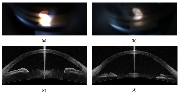
Purpose. To evaluate interobserver, intervisit, and interinstrument agreements for gonioscopy and Fourier domain anterior segment optical coherence tomography (FD ASOCT) for classifying open and narrow angle eyes. Methods. Eighty-six eyes with open or narrow anterior chamber angles were included. The superior angle was classified open or narrow by 2 of 5 glaucoma specialists using gonioscopy and imaged by FD ASOCT in the dark. The superior angle of each FD ASOCT image was graded as open or narrow by 2 masked readers. The same procedures were repeated within 6 months. Kappas for interobserver and intervisit agreements for each instrument and interinstrument agreements were calculated. Results. The mean age was 50.9 (±18.4) years. Interobserver agreements were moderate to good for both gonioscopy (0.57 and 0.69) and FD ASOCT (0.58 and 0.75). Intervisit agreements were moderate to excellent for both gonioscopy (0.53 to 0.86) and FD ASOCT (0.57 and 0.85). Interinstrument agreements were fair to good (0.34 to 0.63), with FD ASOCT classifying more angles as narrow than gonioscopy. Conclusions. Both gonioscopy and FD ASOCT examiners were internally consistent with similar interobserver and intervisit agreements for angle classification. Agreement between instruments was fair to good, with FD ASOCT classifying more angles as narrow than gonioscopy.
Conflict of interest statement
No authors declare any relevant conflict of interests.
Figures
Similar articles
-
Comparison of EyeCam and anterior segment optical coherence tomography in detecting angle closure.Acta Ophthalmol. 2012 Dec;90(8):e621-5. doi: 10.1111/j.1755-3768.2012.02510.x. Epub 2012 Aug 31. Acta Ophthalmol. 2012. PMID: 22938754
-
Evaluation of the Anterior Segment Angle-to-Angle Scan of Cirrus High-Definition Optical Coherence Tomography and Comparison With Gonioscopy and With the Visante OCT.Invest Ophthalmol Vis Sci. 2017 Jan 1;58(1):59-64. doi: 10.1167/iovs.16-20886. Invest Ophthalmol Vis Sci. 2017. PMID: 28061511
-
Classification algorithms based on anterior segment optical coherence tomography measurements for detection of angle closure.Ophthalmology. 2013 Jan;120(1):48-54. doi: 10.1016/j.ophtha.2012.07.005. Epub 2012 Sep 23. Ophthalmology. 2013. PMID: 23009888
-
Clinical role of swept source optical coherence tomography in anterior segment diseases: a review.Semin Ophthalmol. 2021 Nov 17;36(8):684-691. doi: 10.1080/08820538.2021.1897854. Epub 2021 Mar 10. Semin Ophthalmol. 2021. PMID: 33689554 Review.
-
Clinical utility of anterior segment swept-source optical coherence tomography in glaucoma.Oman J Ophthalmol. 2016 Jan-Apr;9(1):3-10. doi: 10.4103/0974-620X.176093. Oman J Ophthalmol. 2016. PMID: 27013821 Free PMC article. Review.
Cited by
-
Anterior Segment Optical Coherence Tomography: Applications for Clinical Care and Scientific Research.Asia Pac J Ophthalmol (Phila). 2019 MarchApril 01;8(2):146-157. doi: 10.22608/APO.201910. Asia Pac J Ophthalmol (Phila). 2019. PMID: 31020820 Free PMC article.
-
Glaucoma Expert-Level Detection of Angle Closure in Goniophotographs With Convolutional Neural Networks: The Chinese American Eye Study.Am J Ophthalmol. 2021 Jun;226:100-107. doi: 10.1016/j.ajo.2021.02.004. Epub 2021 Feb 9. Am J Ophthalmol. 2021. PMID: 33577791 Free PMC article.
-
Comparison of the efficacy and invasiveness of manual and automated gonioscopy.PLoS One. 2023 Apr 6;18(4):e0284098. doi: 10.1371/journal.pone.0284098. eCollection 2023. PLoS One. 2023. PMID: 37023115 Free PMC article.
-
Changes in ocular morphology after cataract surgery in open angle glaucoma patients.Sci Rep. 2021 Jun 9;11(1):12203. doi: 10.1038/s41598-021-91740-z. Sci Rep. 2021. PMID: 34108591 Free PMC article.
-
Changes in anterior chamber biometry and intraocular pressure after uneventful phacoemulsification in non-glaucomatous eyes.Med Hypothesis Discov Innov Ophthalmol. 2023 May 31;12(1):28-35. doi: 10.51329/mehdiophthal1467. eCollection 2023 Spring. Med Hypothesis Discov Innov Ophthalmol. 2023. PMID: 37641670 Free PMC article.
References
Grants and funding
LinkOut - more resources
Full Text Sources
Other Literature Sources
