. 2016 Jul 7;2016(6):39-55.
doi: 10.2147/rrbc.s107070.

Identification of Human Cathelicidin Peptide LL-37 as a Ligand for Macrophage Integrin αMβ2 (Mac-1, CD11b/CD18) that Promotes Phagocytosis by Opsonizing Bacteria

Affiliations

Identification of Human Cathelicidin Peptide LL-37 as a Ligand for Macrophage Integrin αMβ2 (Mac-1, CD11b/CD18) that Promotes Phagocytosis by Opsonizing Bacteria

Valeryi K Lishko et al. Res Rep Biochem. .

Abstract

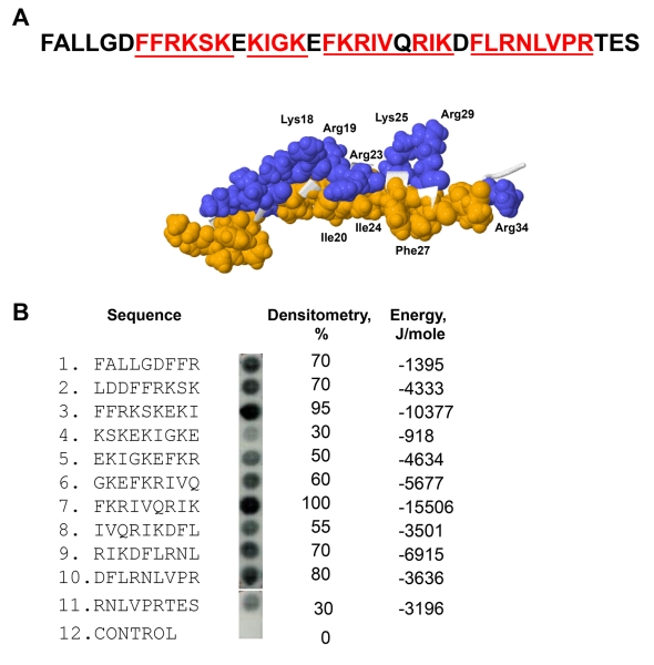
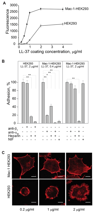
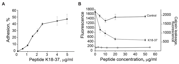
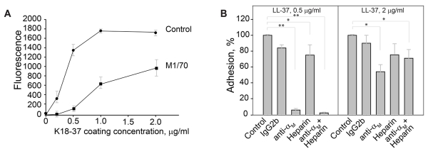
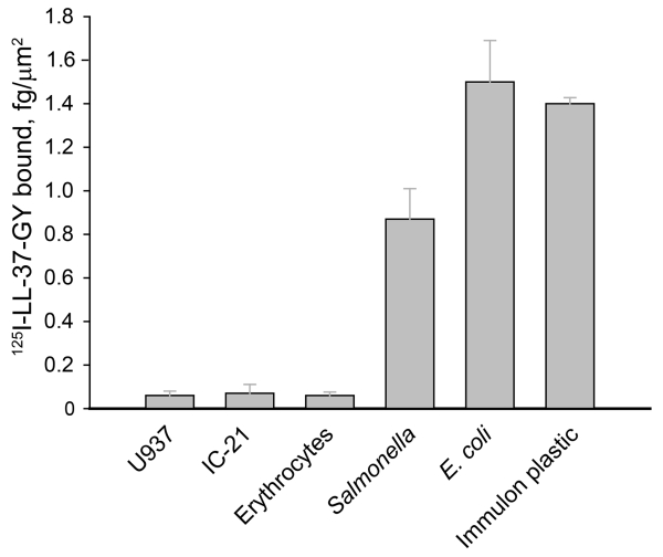
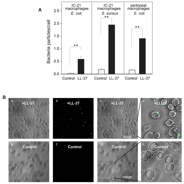
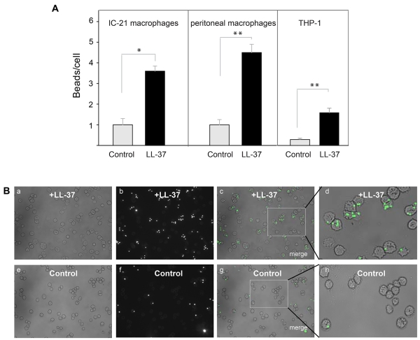
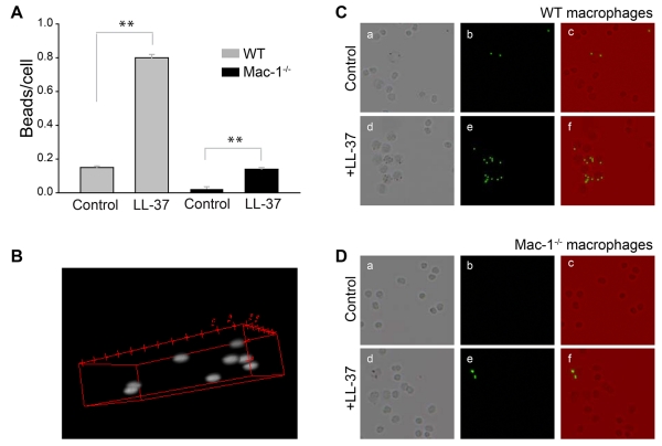
LL-37, a cationic antimicrobial peptide, has numerous immune-modulating effects. However, the identity of a receptor(s) mediating the responses in immune cells remains uncertain. We have recently demonstrated that LL-37 interacts with the αMI-domain of integrin αMβ2 (Mac-1), a major receptor on the surface of myeloid cells, and induces a migratory response in Mac-1-expressing monocyte/macrophages as well as activation of Mac-1 on neutrophils. Here, we show that LL-37 and its C-terminal derivative supported strong adhesion of various Mac-1-expressing cells, including HEK293 cells stably transfected with Mac-1, human U937 monocytic cells and murine IC-21 macrophages. The cell adhesion to LL-37 was partially inhibited by specific Mac-1 antagonists, including mAb against the αM integrin subunit and neutrophil inhibitory factor, and completely blocked when anti-Mac-1 antibodies were combined with heparin, suggesting that cell surface heparan sulfate proteoglycans act cooperatively with integrin Mac-1. Coating both Gram-negative and Gram-positive bacteria with LL-37 significantly potentiated their phagocytosis by macrophages, and this process was blocked by a combination of anti-Mac-1 mAb and heparin. Furthermore, phagocytosis by wild-type murine peritoneal macrophages of LL-37-coated latex beads, a model of foreign surfaces, was several fold higher than that of untreated beads. By contrast, LL-37 failed to augment phagocytosis of beads by Mac-1-deficient macrophages. These results identify LL-37 as a novel ligand for integrin Mac-1 and demonstrate that the interaction between Mac-1 on macrophages and bacteria-bound LL-37 promotes phagocytosis.

Keywords: CD11b/CD18; LL-37; Mac-1; integrin αMβ2; opsonin; phagocytosis.


Figures

Figure 1
Figure 1. Characterization of the αMI-domain recognition motifs in the FALL-39 sequence
(A) The amino acid sequence of FALL-39 and the 3D structure of LL-37 based on PDB Id: 2K6O. Positively charged (blue) and hydrophobic (tan) residues in the C-terminal part of the peptide are numbered. The underlined sequences denote the αMI-domain recognition patterns. (B) The peptide library derived from the FALL-39 sequence (left panel) consisting of 9-mer peptides with a 3 residue offset was incubated with 125I-labeled αMI-domain and the αMI-domain binding was visualized by autoradiography. Control, a spot containing only the β-Ala spacer used for the attachment of peptides to the cellulose membrane. The αMI-domain binding observed as dark spots was analyzed by densitometry (middle column). The numbers show the relative binding of the αMI-domain to peptides expressed as a percentage of the intensity of spot 3. The peptide energies (right column) that serve as a measure of probability each peptide can interact with the αMI-domain were calculated as described.
Figure 2
Figure 2. LL-37 supports adhesion of the αMβ2-expresing cells
(A) Aliquots (100 μl; 5×10⁴/ml) of Mac-1-expressing HEK293 (Mac-1-HEK293) and wild-type HEK293 (HEK293) cells labeled with calcein were added to microtiter wells coated with different concentrations of LL-37 and postcoated with 1% PVP. After 30 min at 37 °C, nonadherent cells were removed by washing and fluorescence of adherent cells was measured in a fluorescence reader. A representative of 6 experiments in which two cell lines were tested side by side is shown. Data presented are means for triplicate determinations, and error bars represent S.E. (B) Wild-type HEK293 cells were preincubated with anti-β1 mAb (10 μg/ml), heparin (20 μg/ml) or their mixture for 15 min at 22 °C and added to wells coated with 2 μg/ml LL-37. Mac-1-HEK293 cells were preincubated with anti-αM mAb 44a (10 μg/ml), heparin (20 μg/ml) of their mixture and added to wells coated with 0.4 or 2 μg/ml LL-37. Adhesion was quantified as described above. Adhesion in the absence of Mac-1 inhibitors and heparin was assigned a value of 100%. Data shown are means ± S.E from 3-4 separate experiments with triplicate measurements. **p≤0.01, *p≤0.05 compared with control adhesion in the absence of inhibitors. (C) Mac-1-HEK293 (upper panel) and HEK293 cells (bottom panel) were plated on glass slides and allowed to adhere for 30 min at 37 °C. Nonadherent cells were removed and adherent cells were fixed with 2% paraformaldehyde followed by staining with Alexa Fluor 546 phalloidin. The cells were imaged with a Leica SP5 laser scanning confocal microscope with a 63× objective.
Figure 3
Figure 3. Adhesion of Mac-1-HEK293 and wild-type HEK293 cells to the LL-37-derived peptide K18-37
(A) Aliquots (100 μl; 5×10⁴/ml) of Mac-1-HEK293 cells were labeled with calcein and added to microtiter wells coated with different concentrations of K18-37. After 30 min at 37 °C, nonadherent cells were removed and adhesion was measured. Adhesion was expressed as percent of added cells. The data shown are means and S.E. from four experiments with triplicate determinations at each point. (B) Calcein-labeled Mac-1-HEK293 cells were incubated with different concentrations of K18-37 (●) or control peptide (▼) for 15 min at 22 °C and added to wells coated with 2.5 μg/ml fibrinogen and post-coated with 1% PVP. Adhesion (left ordinate) was determined as described in Fig. 2A legend. Right ordinate, Mac-1-HEK293 cells were treated with different concentrations of K18-37 for 15 min at 22 °C. Cells were centrifuged and fluorescence of cell supernatants determined (○). The data shown are the mean ± S.E. from two experiments each with triplicate determinations.
Figure 4
Figure 4. Adhesion of murine IC-21 macrophages to the LL-37-derived peptide K18-37
(A) Calcein-labeled IC-21 macrophages were preincubated for 15 min at 22 °C with buffer (●) or 10 μg/ml mAb M1/70 (anti-αM) (■). Aliquots (5×10⁴/0.1 ml) of cells were added to microtiter wells coated with different concentrations of K18-37 (0-2 μg/ml) and postcoated with 1% PVP. After 30 min at 37 °C, nonadherent cells were removed by washing and adhesion was determined. (B) IC-21 macrophages were preincubated with anti-αM mAb M1/70 (10 μg/ml), heparin (20 μg/ml) or their mixture and added to wells coated with 0.5 and 2 μg/ml K18-37. Adhesion was quantified as described above. Adhesion in the absence of inhibitors and heparin was assigned a value of 100 %. The data shown are the mean ± S.E. from 5-6 experiments each with triplicate determinations. **p≤0.01 and *p≤0.05 compared with control adhesion in the absence of inhibitors.
Figure 5
Figure 5. Binding density of LL-37 on the surface of mammalian and microbial cells
Cell suspensions were incubated with 5 μg/ml 125I-LL-37-GY for 20 min at 22 °C. Non-bound peptide was removed by centrifugation at 200g (mammalian cells) or 2000g (bacteria) for 10 min. The pellet was washed with PBS, re-suspended in 100 μl PBS and radioactivity was measured. The adsorption of 125I-LL-37-GY to Immulon wells is described in the legend to Supplemental Fig. 1. The data shown are the mean ± S.E. from 3 experiments each with duplicate measurements.
Figure 6
Figure 6. Effect of LL-37 on phagocytosis of S. aureus particles and E. coli by suspensions of murine IC-21 macrophages
(A) Fluorescent S. aureus particles (4×10⁷/ml) were preincubated with LL-37 (5 μg/ml), K18-37 (10 μg/ml) or polyL-lysine (10 μg/ml) for 20 min at 22 °C. Soluble peptide was removed by centrifugation. Peptide-coated particles were incubated with suspensions of IC-21 macrophages (106/ml) for 60 min at 37 °C and nonphagocytosed particles were separated from cells by filtering the suspensions using a 3-μm pore Transwell inserts. Macrophages were transferred to wells of 96-well plates and trypan blue was added to wells. The ratio of bacterial particles per macrophage was quantified for five random fields per well using a 20× objective. Data shown are mean/cell ± S.E. from five or more experiments. **p≤0.01, compared with untreated control S. aureus. (B) Bright field (a,e), fluorescence (b,f) and merged (c,g) images of IC-21 macrophages incubated with LL-37-coated (a-c) or uncoated (e-g) control bacteria. The representative low power (20×) fields are shown. Enlarged images (d,h) of macrophages shown in the boxed areas in c and g. Phagocytosed bacterial particles are labeled in green. (C) Concentration-dependent effect of LL-37 on phagocytosis of E. coli by macrophages. Fluorescently labeled E. coli cells were incubated with different concentrations of LL-37 for 20 min at 22 °C. After the removal of nonbound peptide by centrifugation, LL-37-coated E. coli were incubated with IC-21 macrophages (106/ml) for 60 min at 37 °C. Phagocytosis was determined as in Fig. 6A. Data are expressed as mean ratios of bacteria per macrophage ± SE. A representative of three experiments is shown. (D) Fluorescent S. aureus particles were preincubated with LL-37 (5 μg/ml) for 20 min at 22 °C. Soluble peptide was removed by centrifugation. Peptide-coated bacterial particles were incubated with IC-21 macrophages (106/ml) in the presence of anti-αM mAb M1/70 (20 μg/ml), heparin (20 μg/ml) or their mixture. After 60 min incubation at 37 °C, nonphagocytosed S. aureus particles were removed and phagocytosis was measured as described above. Data shown are mean bacterial particles/cell ± S.E. of five random fields determined for each condition and are representative of 3 separate experiments. **p≤0.01, *p≤0.05
Figure 7
Figure 7. Augmentation of phagocytosis of LL-37-coated bacteria by adherent macrophages
(A) Fluorescently labeled S. aureus particles and E. coli cells were incubated with LL-37 (5 μg/ml) for 20 min at 22 °C. Soluble peptide was removed by centrifugation and LL-37-coated bacteria were subsequently incubated with adherent IC-21 macrophages or mouse peritoneal macrophages for 60 min at 37 °C. Data are expressed as mean ratios of bacteria per macrophage ± SE. The panels shown in A are representative of three or more experiments. **p≤0.01 compared with untreated control bacteria. (B) A representative experiment showing bright field (a,e), fluorescence (b,f) and merged images (c,g) of IC-21 macrophages exposed to LL-37-coated and uncoated control S. aureus.
Figure 8
Figure 8. Effect of LL-37 on phagocytosis of latex beads
(A) Fluorescent latex beads (2.5×10⁷/ml) were preincubated with LL-37 (10 μg/ml) for 20 min at 22 °C. Soluble peptide was removed from beads by high-speed centrifugation. Peptide-coated beads were incubated with IC-21 cells, mouse peritoneal macrophages or differentiated THP-1 cells for 30 min at 37 °C. Nonphagocytosed beads were separated from macrophages by centrifugation. The ratio of beads per macrophage was quantified from three fields of fluorescent images. Data shown are means ± S.E. of triplicate measurements and are representative of 3 experiments. **p≤0.01, *p≤0.05 compared with untreated beads. (B) Fluorescence of IC-21 macrophages exposed to LL-37-treated beads (a,b,c) or control untreated beads (e,f,g). Enlarged images of IC-21 macrophages showing phagocytosed LL-37-treated (d) or control (h) beads.
Figure 9
Figure 9. Effect of LL-37 on phagocytosis of latex beads by Mac-1-deficient macrophages
(A) Resident peritoneal macrophages were obtained from WT and Mac-1−/− mice as described in the Materials and Methods. LL-37-coated beads (prepared as in Fig. 8) were incubated with adherent macrophages for 30 min at 37 °C. Nonphagocytosed beads were washed, cells treated with trypan blue and the ratio of beads per macrophage was quantified from three fields of fluorescent images. Data shown are means ± S.E. of triplicate measurements from three experiments. **p≤0.01 compared with untreated control beads. (B) Representative confocal image illustrates phagocytosed beads inside a macrophage. (C, D) The representative fields of WT (C) and Mac-1-deficient (D) macrophages incubated with control and LL-37-coated beads. Bright field (a,d), fluorescence (b,e) and merged (c,f) images of WT and Mac-1-deficient macrophages.

Similar articles

Cited by

References

    1. Zanetti M. The role of cathelicidins in the innate host defenses of mammals. In: Gallo RL, editor. Antimicrobial peptides in human health and disease. Horizon Bioscience; Norfolk, U.K.: 2005. pp. 15–50.
    1. Sorensen OE, Follin P, Johnsen AH, et al. Human cathelicidin, hCAP-18, is processed to the antimicrobial peptide LL-37 by extracellular cleavage with proteinase 3. Blood. 2001;97:3951–3959. - PubMed
    1. Gundmundsson GH, Agerberth B. Antimicrobial peptides in human blood. In: Gallo RL, editor. Antimicrobial peptides in human health and disease. Horizon Bioscience; Norfolk, U.K.: 2005. pp. 153–174.
    1. Bals R, Weiner DJ, Moscioni AD, Meegalla RL, Wilson JM. Augmentation of innate host defense by expression of a cathelicidin antimicrobial peptide. Infect Immun. 1999;67:6084–6089. - PMC - PubMed
    1. Cirioni O, Giacometti A, Ghiselli R, et al. LL-37 protects rats against lethal sepsis caused by gram-negative bacteria. Antimicrob Agents Chemother. 2006;50:1672–1679. - PMC - PubMed

LinkOut - more resources