

Mutations in the Human AAA+ Chaperone p97 and Related Diseases
- PMID: 27990419
- PMCID: PMC5131264
- DOI: 10.3389/fmolb.2016.00079
Mutations in the Human AAA+ Chaperone p97 and Related Diseases
Abstract
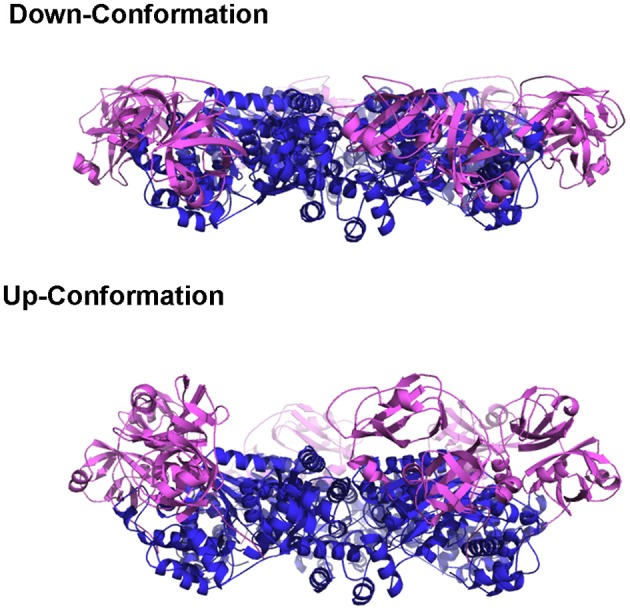
A number of neurodegenerative diseases have been linked to mutations in the human protein p97, an abundant cytosolic AAA+ (ATPase associated with various cellular activities) ATPase, that functions in a large number of cellular pathways. With the assistance of a variety of cofactors and adaptor proteins, p97 couples the energy of ATP hydrolysis to conformational changes that are necessary for its function. Disease-linked mutations, which are found at the interface between two main domains of p97, have been shown to alter the function of the protein, although the pathogenic mutations do not appear to alter the structure of individual subunit of p97 or the formation of the hexameric biological unit. While exactly how pathogenic mutations alter the cellular function of p97 remains unknown, functional, biochemical and structural differences between wild-type and pathogenic mutants of p97 are being identified. Here, we summarize recent progress in the study of p97 pathogenic mutants.
Keywords: VCP/p97; conformational changes; multisystem diseases; mutations; structure and function.
Figures


Similar articles
-
A Mighty "Protein Extractor" of the Cell: Structure and Function of the p97/CDC48 ATPase.Front Mol Biosci. 2017 Jun 13;4:39. doi: 10.3389/fmolb.2017.00039. eCollection 2017. Front Mol Biosci. 2017. PMID: 28660197 Free PMC article. Review.
-
Molecular Mechanisms Driving and Regulating the AAA+ ATPase VCP/p97, an Important Therapeutic Target for Treating Cancer, Neurological and Infectious Diseases.Biomolecules. 2023 Apr 24;13(5):737. doi: 10.3390/biom13050737. Biomolecules. 2023. PMID: 37238606 Free PMC article. Review.
-
Molecular perspectives on p97-VCP: progress in understanding its structure and diverse biological functions.J Struct Biol. 2004 Apr-May;146(1-2):44-57. doi: 10.1016/j.jsb.2003.11.014. J Struct Biol. 2004. PMID: 15037236 Review.
-
High-speed atomic force microscopic observation of ATP-dependent rotation of the AAA+ chaperone p97.Structure. 2013 Nov 5;21(11):1992-2002. doi: 10.1016/j.str.2013.08.017. Epub 2013 Sep 19. Structure. 2013. PMID: 24055316
-
The Interplay of Cofactor Interactions and Post-translational Modifications in the Regulation of the AAA+ ATPase p97.Front Mol Biosci. 2017 Apr 13;4:21. doi: 10.3389/fmolb.2017.00021. eCollection 2017. Front Mol Biosci. 2017. PMID: 28451587 Free PMC article. Review.
Cited by
-
Fragment screening using biolayer interferometry reveals ligands targeting the SHP-motif binding site of the AAA+ ATPase p97.Commun Chem. 2022 Dec 7;5(1):169. doi: 10.1038/s42004-022-00782-5. Commun Chem. 2022. PMID: 36697690 Free PMC article.
-
SVIP is a molecular determinant of lysosomal dynamic stability, neurodegeneration and lifespan.Nat Commun. 2021 Jan 21;12(1):513. doi: 10.1038/s41467-020-20796-8. Nat Commun. 2021. PMID: 33479240 Free PMC article.
-
The AAA + ATPase valosin-containing protein (VCP)/p97/Cdc48 interaction network in Leishmania.Sci Rep. 2020 Aug 4;10(1):13135. doi: 10.1038/s41598-020-70010-4. Sci Rep. 2020. PMID: 32753747 Free PMC article.
-
Phospho-Ser784-VCP Is Required for DNA Damage Response and Is Associated with Poor Prognosis of Chemotherapy-Treated Breast Cancer.Cell Rep. 2020 Jun 9;31(10):107745. doi: 10.1016/j.celrep.2020.107745. Cell Rep. 2020. PMID: 32521270 Free PMC article.
-
Host AAA+ ATPase TER94 Plays Critical Roles in Building the Baculovirus Viral Replication Factory and Virion Morphogenesis.J Virol. 2020 Feb 28;94(6):e01674-19. doi: 10.1128/JVI.01674-19. Print 2020 Feb 28. J Virol. 2020. PMID: 31896597 Free PMC article.
References
Publication types
LinkOut - more resources
Full Text Sources
Other Literature Sources
Miscellaneous