. 2016:2016:7616471.
doi: 10.1155/2016/7616471. Epub 2016 Nov 20.

Enhanced Performance and Mode of Action of a Novel Antibiofilm Hydrofiber® Wound Dressing

Affiliations

Enhanced Performance and Mode of Action of a Novel Antibiofilm Hydrofiber® Wound Dressing

David Parsons et al. Biomed Res Int. 2016.

Abstract

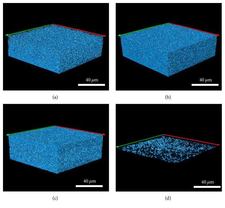
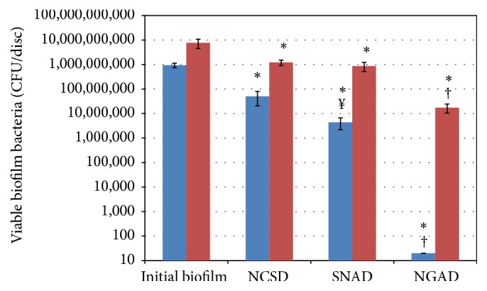
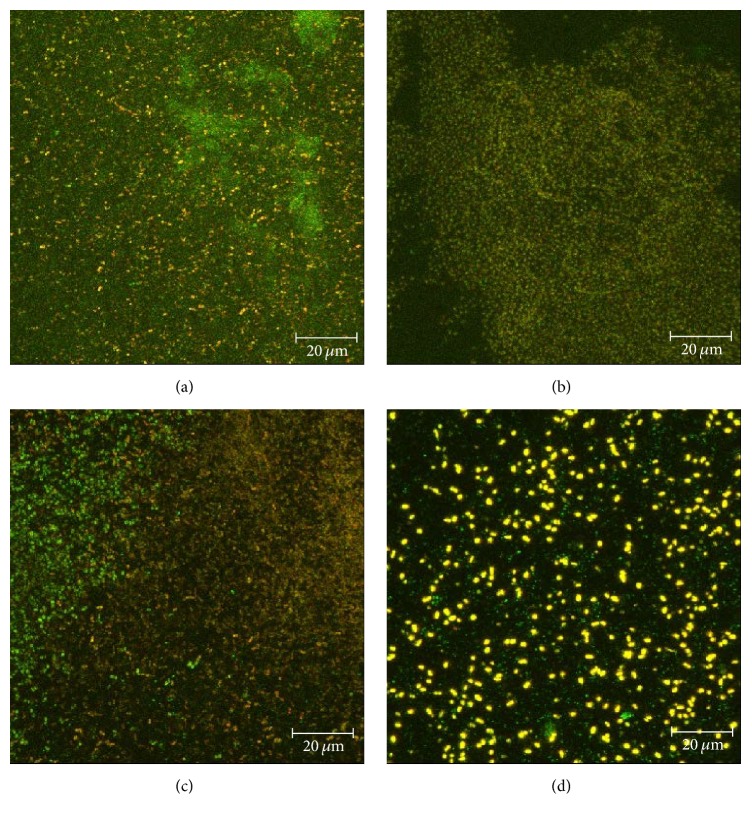
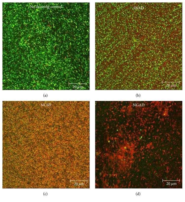
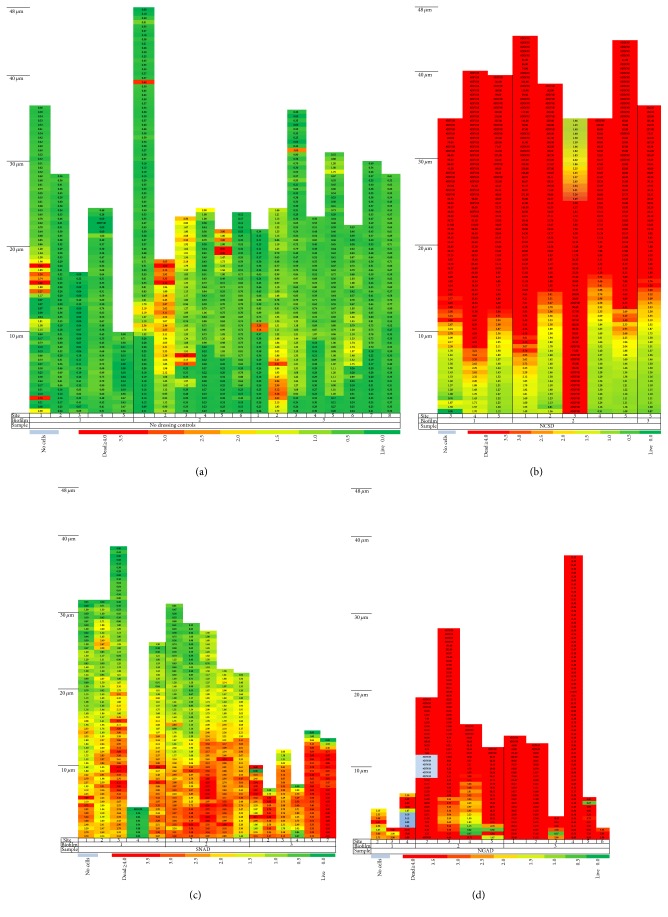
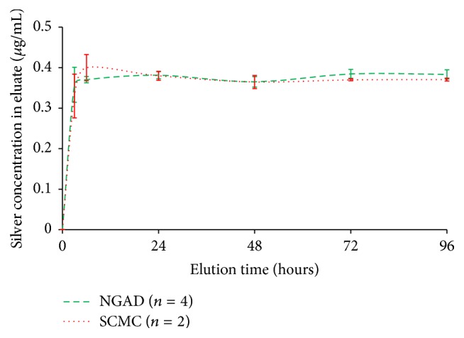
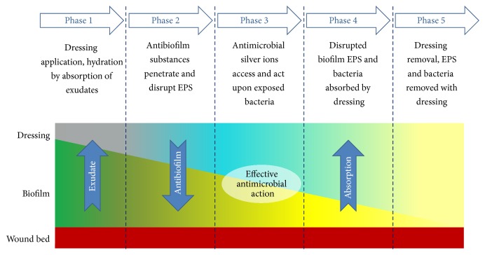
Biofilm development in wounds is now acknowledged to be a precursor to infection and a cause of delayed healing. A next-generation antibiofilm carboxymethylcellulose silver-containing wound dressing (NGAD) has been developed to disrupt and kill biofilm microorganisms. This in vitro study aimed to compare its effectiveness against various existing wound dressings and examine its mode of action. A number of biofilm models of increasing complexity were used to culture biofilms of wound-relevant pathogens, before exposure to test dressings. Confocal microscopy, staining, and imaging of biofilm constituents, total viable counting, and elemental analysis were conducted to assess dressing antibiofilm performance. Live/dead staining and viable counting of biofilms demonstrated that the NGAD was more effective at killing biofilm bacteria than two other standard silver dressings. Staining of biofilm polysaccharides showed that the NGAD was also more effective at reducing this protective biofilm component than standard silver dressings, and image analyses confirmed the superior biofilm killing and removal performance of the NGAD. The biofilm-disruptive and silver-enhancing modes of action of the NGAD were supported by significant differences (p < 0.05) in biofilm elemental markers and silver donation. This in vitro study improves our understanding of how antibiofilm dressing technology can be effective against the challenge of biofilm.


Conflict of interest statement

The authors declare that they have no competing interests.

Figures

Figure 1
Figure 1
(a) 24-hour S. aureus biofilm supported on a 25 mm filter disc in contact with BGM in a deep 6-well plate (biofilm is stained pink for clarity). (b) Test dressing application to biofilm (biofilm is stained pink for clarity).
Figure 2
Figure 2
Simulated wound polymicrobial biofilm model with the NGAD and AQUACEL Foam secondary dressing application within the wound assembly.
Figure 3
Figure 3
Representative isosurface 3D imaging, performed using the Image-Pro Premier® 3D software of the CLSM images biofilms stained with BacLight® (green = viable bacteria; red = nonviable bacteria). (a) Initial biofilm T 24 hours. (b) NCSD after 48 hours of exposure. (c) SNAD after 48 hours of exposure. (d) NGAD after 48 hours of exposure.
Figure 4
Figure 4
Percentage of EPS still remaining after exposure to the silver test dressings for 48 hours. p = 0.000.
Figure 5
Figure 5
Representative isosurface 3D imaging, performed using the Image-Pro Premier 3D software, of the CLSM images biofilm EPS stained with Calcofluor White. (a) Initial biofilm T 24 hours. (b) NCSD after 48 hours of exposure. (c) SNAD after 48 hours of exposure. (d) NGAD after 48 hours of exposure.
Figure 6
Figure 6
Total viable counts of (blue colour) S. aureus and (red colour) K. pneumoniae biofilm cells after 48 hours of exposure to silver dressings (n = 5). Initial biofilm = T 24 hours. p < 0.05 compared to initial biofilm. p = 0.000 compared to NCSD and SNAD. ¥ p = 0.027 compared to NCSD.
Figure 7
Figure 7
Representative CLSM images of polymicrobial biofilm where bacteria have been fluorescently tagged (green = S. aureus; yellow = K. pneumoniae). (a) Initial biofilm T 24 hours; (b) NSCD after 48 hours; (c) SNAD after 48 hours; (d) NGAD after 48 hours.
Figure 8
Figure 8
Composite (full thickness) images of antibiotic-resistant P. aeruginosa biofilm stained with BacLight after 48 hours of contact with the test dressings (green = viable bacteria; red = nonviable bacteria). (a) No-dressing control. (b) SNAD. (c) NCSD. (d) NGAD.
Figure 9
Figure 9
Colour-coded bacterial viability within biofilm layers as a function of distance from the filter surface for an antibiotic-resistant P. aeruginosa biofilm after 48 hours of contact with the test dressings (green = viable bacteria; red = nonviable bacteria). (a) No-dressing control. (b) NCSD. (c) SNAD. (d) NGAD.
Figure 10
Figure 10
Comparison of elution of silver ions from the NGAD and SCMC into a constantly stirred excess of isotonic media (0.9% w/v NaCl(aq), 8 mL per cm2 dressing at 37 ± 3°C) as determined by ICP-MS.
Figure 11
Figure 11
Functionality of the NGAD.

Similar articles

Cited by

References

    1. Bowler P. G., Welsby S., Towers V., et al. Multidrug-resistant organisms, wounds and topical antimicrobial protection. International Wound Journal. 2012;9(4):387–396. doi: 10.1111/j.1742-481x.2012.00991.x. - DOI - PMC - PubMed
    1. Bowler P., Jones S., Towers V., Booth R., Parsons D., Walker M. Dressing conformability and silver-containing wound dressings. Wounds UK. 2010;6(2):14–20.
    1. Flemming H.-C., Wingender J. The biofilm matrix. Nature Reviews Microbiology. 2010;8(9):623–633. doi: 10.1038/nrmicro2415. - DOI - PubMed
    1. James G. A., Swogger E., Wolcott R., et al. Biofilms in chronic wounds. Wound Repair and Regeneration. 2008;16(1):37–44. doi: 10.1111/j.1524-475x.2007.00321.x. - DOI - PubMed
    1. Kirketerp-Møller K., Jensen P. Ø., Fazli M., et al. Distribution, organization, and ecology of bacteria in chronic wounds. Journal of Clinical Microbiology. 2008;46(8):2717–2722. doi: 10.1128/JCM.00501-08. - DOI - PMC - PubMed

MeSH terms