

Low-dose lung cancer screening with photon-counting CT: a feasibility study
- PMID: 27991453
- PMCID: PMC5237389
- DOI: 10.1088/1361-6560/62/1/202
Low-dose lung cancer screening with photon-counting CT: a feasibility study
Abstract
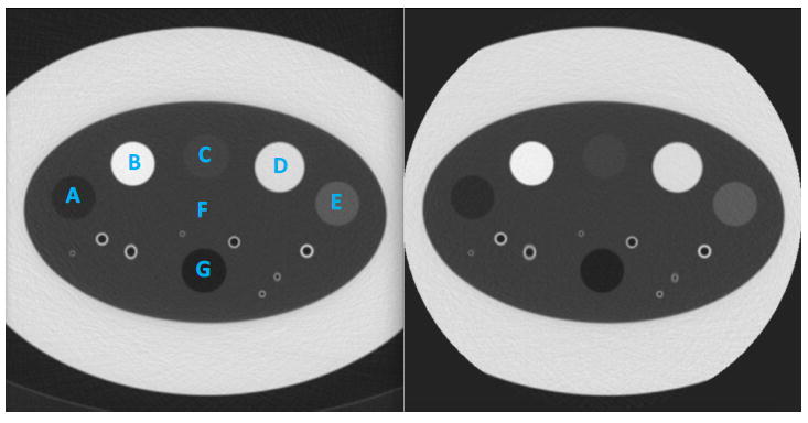
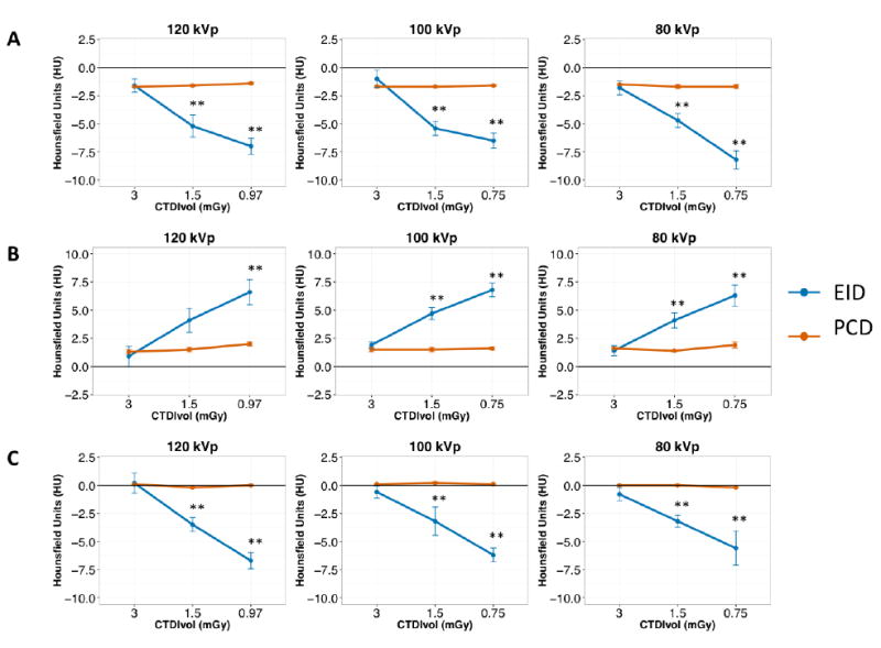
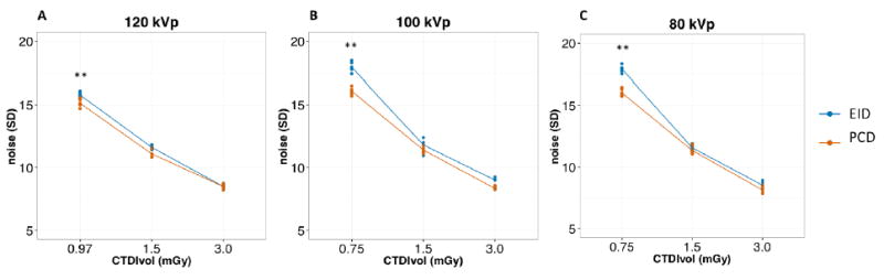
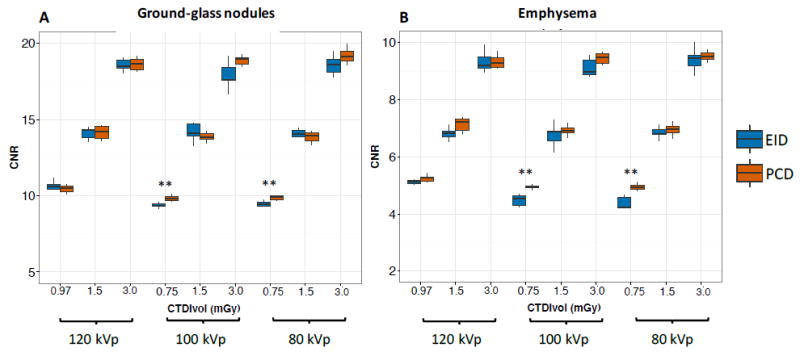
To evaluate the feasibility of using a whole-body photon-counting detector (PCD) CT scanner for low-dose lung cancer screening compared to a conventional energy integrating detector (EID) system. Radiation dose-matched EID and PCD scans of the COPDGene 2 phantom were acquired at different radiation dose levels (CTDIvol: 3.0, 1.5, and 0.75 mGy) and different tube voltages (120, 100, and 80 kVp). EID and PCD images were compared for quantitative Hounsfield unit (HU) accuracy, noise levels, and contrast-to-noise ratios (CNR) for detection of ground-glass nodules (GGN) and emphysema. The PCD HU accuracy was better than EID for water at all scan parameters. PCD HU stability for lung, GGN and emphysema regions were superior to EID and PCD attenuation values were more reproducible than EID for all scan parameters (all P < 0.01), while HUs for lung, GGN and emphysema ROIs changed significantly for EID with decreasing dose (all P < 0.001). PCD showed lower noise levels at the lowest dose setting at 120, 100 and 80 kVp (15.2 ± 0.3 HU versus 15.8 ± 0.2 HU, P = 0.03; 16.1 ± 0.3 HU versus 18.0 ± 0.4 HU, P = 0.003; and 16.1 ± 0.3 HU versus 17.9 ± 0.3 HU, P = 0.001, respectively), resulting in superior CNR for evaluation of GGNs and emphysema at 100 and 80 kVp. PCD provided better HU stability for lung, ground-glass, and emphysema-equivalent foams at lower radiation dose settings with better reproducibility than EID. Additionally, PCD showed up to 10% less noise, and 11% higher CNR at 0.75 mGy for both 100 and 80 kVp. PCD technology may help reduce radiation exposure in lung cancer screening while maintaining diagnostic quality.
Conflict of interest statement
Authors who are not employees of or consultants for Siemens had control of inclusion of any data and information that might present a conflict of interest for the authors who are employed by Siemens.
Figures
References
-
- Alvarez RE, Macovski A. Energy-selective reconstructions in x-ray computerised tomography. Phys Med Biol. 1976;21:733. - PubMed
-
- Baek J, Pineda AR, Pelc NJ. To bin or not to bin? The effect of CT system limiting resolution on noise and detectability. Phys Med Biol. 2013;58:1433. - PubMed
-
- Dixon RL. A new look at CT dose measurement: beyond CTDI. Med Phys. 2003;30:1272–80. - PubMed
-
- Funama Y, Awai K, Liu D, Oda S, Yanaga Y, Nakaura T, Kawanaka K, Shimamura M, Yamashita Y. Detection of nodules showing ground-glass opacity in the lungs at low-dose multidetector computed tomography: phantom and clinical study. J Comput Assist Tomogr. 2009;33:49–53. - PubMed
MeSH terms
Grants and funding
LinkOut - more resources
Full Text Sources
Other Literature Sources
Medical