. 2016 Dec 19:6:39010.
doi: 10.1038/srep39010.

The Identification and Characterization of Two Novel Epitopes on the Nucleocapsid Protein of the Porcine Epidemic Diarrhea Virus

Affiliations

The Identification and Characterization of Two Novel Epitopes on the Nucleocapsid Protein of the Porcine Epidemic Diarrhea Virus

Kang Wang et al. Sci Rep. .

Abstract

Porcine epidemic diarrhea virus (PEDV) is a highly contagious coronavirus that causes severe diarrhea and death, particularly in neonatal piglets. The nucleocapsid protein (N protein) of PEDV presents strong immunogenicity and contributes to the cross-reactivity between PEDV and TGEV. However, the characterization of epitopes on the PEDV N protein remains largely unknown. Here, two monoclonal antibodies (MAbs) specific to the N protein of a PEDV strain, FJzz1/2011, were generated and screened against a partially overlapping library of 24 GST-fusion N protein-truncated constructs. We confirmed that residues 18-133 (designated NEP-D4) and residues 252-262 (designated NEP-D6) were the epitopes targeted by MAbs PN-D4 and PN-D6, respectively. Sequence analysis revealed that these two epitopes were highly conserved among PEDV strains but were significantly different from other members of the Coronavirinae subfamily. Western blot analysis showed that they could be specifically recognized by PEDV antisera but could not be recognized by TGEV hyperimmune antisera. Indirect immunofluorescence (IFA) assays confirmed no cross-reaction between these two MAbs and TGEV. In addition, the freeze-thaw cycle and protease treatment results indicated that NEP-D4 was intrinsically disordered. All these results suggest that these two novel epitopes and their cognate MAbs could serve as the basis for the development of precise diagnostic assays for PEDV.


Conflict of interest statement

The authors declare no competing financial interests.

Figures

Figure 1
Figure 1. Selection of MAbs against PEDV N protein.
(a) Six MAbs were proven to be positive by IFA using PEDV inoculated cells. (b) The six positive clones were then tested by ELISA against GST-fusion N protein and GST-tag expressed in Escherichia coli.
Figure 2
Figure 2. Mapping of PEDV N protein epitopes.
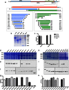
(a) Schematic diagram of the epitope mapping. The segments that could be recognized by both PN-D4 and PN-D6 are highlighted in red; the segments that could only be recognized by PN-D4 are highlighted in blue; the segments that could only be recognized by PN-D6 are highlighted in green; and the segments in gray are those that could not be recognized by either PN-D4 or PN-D6. According to (a), the N gene was divided into mutually overlapping N1 and N2 fragments and was directionally cloned into the pGEX-6p-1 vector. (b) Six hours after the addition of 1 mM IPTG, the induced products of GST-N, GST-N1 and GST-N2 were processed and analyzed using SDS-PAGE (12% separating gel and 5% stacking gel). (c) Next, the GST fusion proteins GST-N, GST-N1 and GST-N2 were coated onto ELISA plates (0.5 μg/well) and were probed with MAbs PN-D4 and PN-D6. After the first round of identification, N1 and N2 were further divided and expressed in two series of fusion proteins, GST-N1-1-GST-N1-11 and GST-N2-1-GST-N2-11 (d,h), which were analyzed by Western blotting and ELISA using PN-D4 (e,g), PN-D6 (i,k), and anti-GST Tag antibody (f,j), respectively.
Figure 3
Figure 3. The reactivity between epitopes and MAbs or antisera.
The two fusion-expressed epitopes (GST-NEP-D4 and GST-NEP-D6) and the negative control (GST tag) were processed and analyzed by Western blotting using PN-D4, PN-D6, or pig PEDV antisera obtained at 14 and 21 days after starting the immunization. Pig TGEV hyperimmune antisera was used as the primary antibodies in membranes a,b,c,d and e. (f) We used the pig TGEV hyperimmune antisera to test GST-TGEV-N protein and GST tag. In IFA, monolayers of Vero cells inoculated with the PEDV strain HLJ/2011 or FJzz1/2011 and ST cells inoculated with the TGEV strain SH/2012 were fixed for IFA against MAbs PN-D4, PN-D6 or anti-TGEV N protein. (g) The PEDV strain HLJ/2011 was detected by MAb PN-D4. (h) The PEDV strain HLJ/2011 was detected by MAb PN-D6. (j) The TGEV strain SH/2012 was detected by MAb PN-D4. (k) The TGEV strain SH/2012 was detected by MAb PN-D6. (i) The PEDV strain FJzz1/2011 was detected by the MAb anti-TGEV N protein. (l) the TGEV strain SH/2012 was detected by the anti-TGEV N protein.
Figure 4
Figure 4. Amino acid sequence analysis of the two identified epitopes among different virus strains.
(a) Amino acid sequence alignments of NEP-D4 and NEP-D6 from 23 PEDV representative strains, including 4 classical strains and 19 currently circulating strains. (b) Similarly, residues 18–133 and residues 252–262 of 21 Alphacoronavirus strains, including seven PEDV strains, six TGEV strains, two human coronavirus 229E strains, two human coronavirus NL63 strains, two PRCV (porcine respiratory coronavirus) strains and two FIPV (feline infectious peritonitis virus) strains, 6 Betacoronavirus strains, including two SARS-CoV (severe acute respiratory syndrome coronavirus) strains, two MERS-CoV (Middle East respiratory syndrome coronavirus) strains and two MHV (mouse hepatitis virus) strains, 2 IBV (Infectious bronchitis virus) strains from Gammacoronavirus, and 3 PDCoV (porcine deltacoronavirus) strains from Deltacoronavirus, were selected for alignment. The corresponding antigenic positions of NEP-D4 and NEP-D6 of all strains are colored green and red, respectively. PEDV strains FJzz1/2011 and HLJ/2011 isolated from our laboratory were marked with black triangles. The major mutations are highlighted by dotted frames.
Figure 5
Figure 5. Identification of IDRs and disulfide bridges in epitope NEP-D4.
(a) The disorder plot for epitope EP4 was predicted by GeneSilico metadisorder. The x-axis shows the residues from 18 to 133, and the y-axis shows the disordered tendency ranging from 0 to 1. All residues with a disorder probability of more than 0.5 were considered to be disordered. The residues with plotted lines in the light blue region were predicted to be disordered; their counterparts in the bluish white area were considered ordered. (b) Four GST-NEP-D4 samples were treated with Trypsin under the same conditions, and the reactions were stopped at 0 min, 1 min, 5 min and 15 min. Next, the samples were processed and analyzed by Western Blotting using anti-GST antibody and PN-D4 as the primary antibodies. (c) Similarly, four additional GST-NEP-D4 samples were treated with freeze (−80 °C) and thaw (room temperature) cycles, which were repeated up to 0, 1, 5 and 10, times, respectively. Next, these samples were analyzed by Western blotting using the same antibodies above. (d) To rule out the interference of disulfide bridges, the reduced (1 M DTT-treated) and non-reduced (non DTT-treated) GST-NEP-D4 were analyzed by Western blotting using PN-D4.
Figure 6
Figure 6. Predicted 3D structural model of N protein with Phyre2.
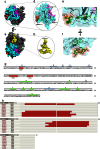
The N protein 3D model was visualized using the PyMOL molecular graphics and modeling system and was viewed from the side (a) and top (c). The protein epitope domains identified in this study are highlighted in different colors. (b) The conformation of the epitope NEP-D6 is displayed with all residues indicated in the corresponding positions. (d) The secondary structure of epitope NEP-D4 is depicted, in which the predicted disordered regions are colored green and tertiary structures showing PIDR1, PIDR2107–116 and PIDR3127–133 are colored blue, magenta and red, respectively. (e,f) The putative spatial positions of critical residues, E133 and L18, which are represented by orange. (g) The sequence of N protein with alpha helixes, beta strands and PIDRs. (h) Template coverage used in the structure modeling.

References

    1. Duarte M., Gelfi J., Lambert P., Rasschaert D. & Laude H. Genome organization of porcine epidemic diarrhoea virus. Adv. Exp. Med. Biol. 342, 55–60 (1993). - PubMed
    1. Enjuanes L., Siddell S. & Spaan W. In Coronaviruses and Arteriviruses (eds Bridgen A., Kocherhans R., Tobler K., Carvajal A. & Ackermann M.) 781–786 (Springer, 1998).
    1. Kocherhans R., Bridgen A., Ackermann M. & Tobler K. Completion of the porcine epidemic diarrhoea coronavirus (PEDV) genome sequence. Virus Genes 23, 137–144 (2001). - PMC - PubMed
    1. Kusanagi K. et al. Isolation and serial propagation of porcine epidemic diarrhea virus in cell cultures and partial characterization of the isolate. J. Vet. Med. Sci. 54, 313–318 (1992). - PubMed
    1. Wood E. N. An apparently new syndrome of porcine epidemic diarrhoea. Vet. Rec. 100, 243–244 (1977). - PubMed

Publication types

MeSH terms