. 2016 Dec 19:6:39429.
doi: 10.1038/srep39429.

A polymorphism of HMGA1 protects against proliferative diabetic retinopathy by impairing HMGA1-induced VEGFA expression

Affiliations

A polymorphism of HMGA1 protects against proliferative diabetic retinopathy by impairing HMGA1-induced VEGFA expression

Eusebio Chiefari et al. Sci Rep. .

Abstract

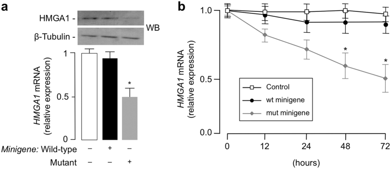
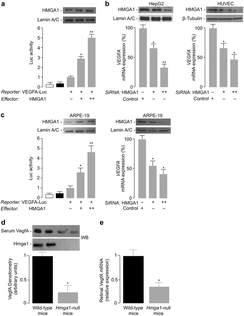
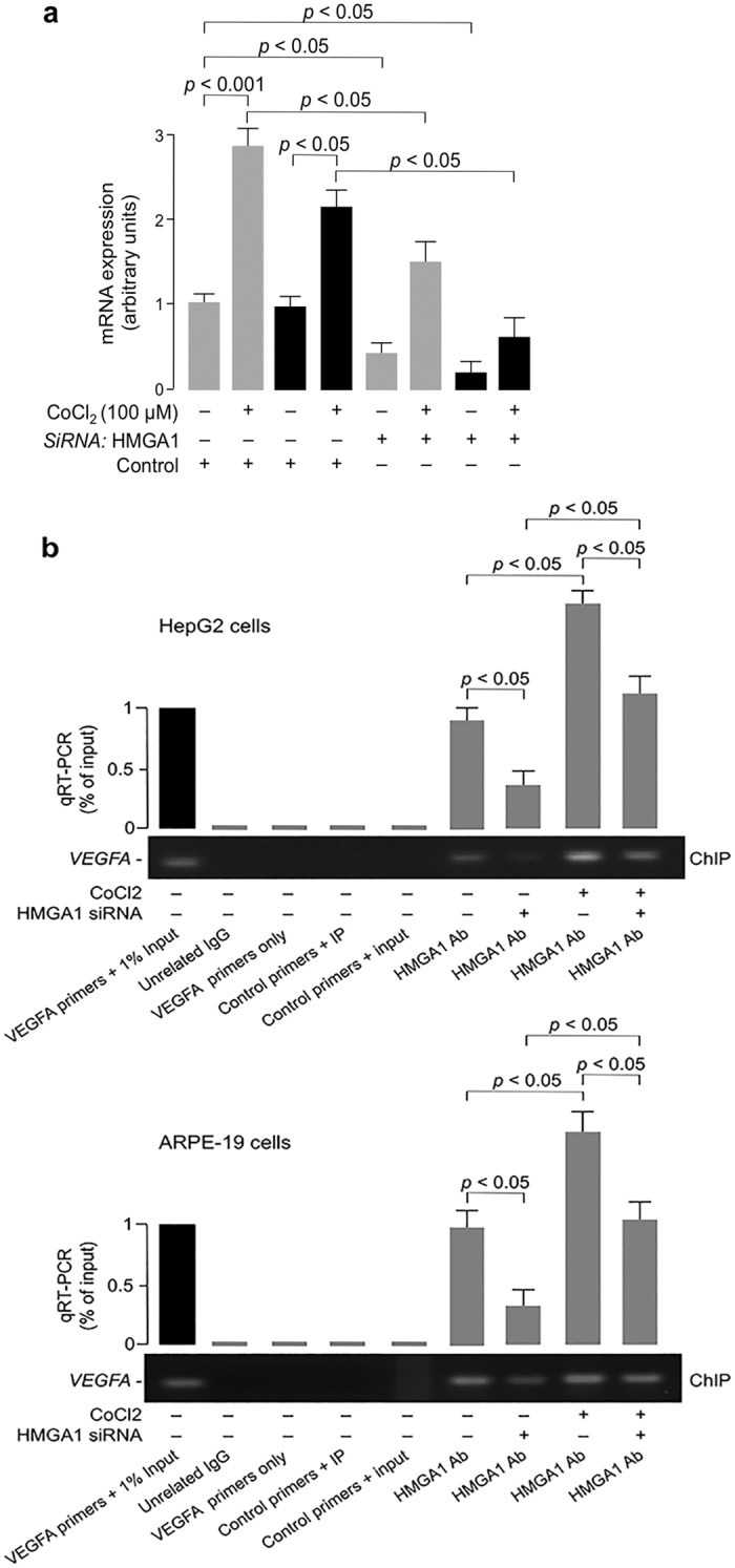
Diabetic retinopathy (DR) is a major complication of diabetes mellitus, and is the leading cause of blindness in working-age people. Usually, DR progresses from the asymptomatic non-proliferative DR that does not significantly alter vision, to proliferative DR (PDR), which can result in aberrant retinal neovessel formation and blindness. The High-Mobility-Group A1 (HMGA1) protein is a transcriptional master regulator of numerous genes, including metabolic and inflammatory genes, which, by modulating the expression of angiogenic factors, may induce retinal neovascularization, a hallmark of PDR. Herein, we examined the relationship between HMGA1 rs139876191 variant and DR. Results revealed that patients with type 2 diabetes, who were carriers of the HMGA1 rs139876191 variant had a significantly lower risk of developing PDR, compared to non-carrier diabetic patients. From a mechanistic point of view, our findings indicated that, by adversely affecting HMGA1 protein expression and function, the HMGA1 rs139876191 variant played a key role in this protective mechanism by downregulating the expression of vascular endothelial growth factor A (VEGFA), a major activator of neovascularization in DR. These data provide new insights into the pathogenesis and progression of DR, and may offer opportunities for discovering novel biomarkers and therapeutic targets for diagnosis, prevention and treatment of PDR.


Figures

Figure 1
Figure 1. Effect of the HMGA1 minigene on HMGA1 expression.
(a) HEK-293 cells were transiently transfected with 1 μg wild-type or mutant minigene construct, and endogenous HMGA1 mRNA and protein levels were measured 72 h later by qRT-PCR and Western blot (WB), respectively. Cropped blots are shown in the figure. Full-length WBs are presented in Supplementary Fig. S1. *P < 0.05 vs control (white bar). β-tubulin, control of protein loading. (b) Time course of HMGA1 mRNA abundance in HEK-293 cells, untreated (control, open square) or treated with wild-type (wt, solid circle) or mutant (mut, solid diamond) HMGA1 minigene. *P < 0.05 vs control. Data are mean ± standard error of the mean (s.e.m) of three independent experiments, each in triplicate.
Figure 2
Figure 2. VEGFA gene expression is induced by HMGA1.
(a) Human VEGFA-Luc reporter vector (2 μg) was transfected into HepG2 cells, in the presence of increasing amounts (0, 0.5, 1 μg) of HMGA1 effector plasmid, and Luc-activity was measured 48 h later. Data represent means ± s.e.m for three separate experiments; values are expressed as the factors by which Luc-activity increased above the level of the activity obtained in transfections with VEGFA-Luc reporter vector plus the empty effector vector (control), which is assigned an arbitrary value of 1. White bar, mock (no DNA); black bar, pGL3-basic (vector without an insert). *P < 0.05 and **P < 0.01 vs control. (b) qRT-PCR of endogenous VEGFA mRNA from HepG2 (left), and HUVEC (right) cells, pretreated with increasing amounts (100 and 200 pmol) of anti-HMGA1 siRNA or nontargeting control siRNA. (c) VEGFA-Luc-activity and qRT-PCR of endogenous VEGFA mRNA were measured in ARPE-19 cells, under the same conditions as in (a) and (b). WBs of HMGA1 in each condition are shown in the autoradiograms. Lamin A/C and β-Tubulin, controls of protein loading. Cropped blots are shown in the figures. Full-length WBs are presented in Supplementary Fig. S1. *P < 0.05 and **P < 0.001 vs siRNA-untreated (control) cells. (d) Representative VegfA WB of blood serum from wild-type and Hmga1-deficient mice. Densitometric analyses of six to eight independent blots are shown. Black bars, wild-type mice, n = 8; gray bars, Hmga1-knockout mice, n = 6. *P < 0.05 vs wild-type controls. Hmga1 protein expression is shown in fat tissue. All the samples were run under the same experimental conditions. Cropped blots are shown in the figures. Full-length WBs are presented in Supplementary Fig. S1. (e) VegfA mRNA levels in retinal tissue of wild-type (black bars) and Hmga1-deficient (gray bars) mice (n = 6 per genotype), as measured by qRT-PCR. Data are means ± s.e.m of three independent measurements from each animal. *P < 0.05 vs wild-type controls.
Figure 3
Figure 3. HMGA1 and VEGFA expression in hypoxia.
(a) Effect of hypoxia on VEGFA (gray bars) and HMGA1 (black bars) mRNA, in HepG2 cells preatreated or not with anti-HMGA1 siRNA, as measured by qRT-PCR. Data are means ± s.e.m of three independent experiments, each performed in triplicate. (b) ChIP of the VEGFA promoter gene in HepG2 and ARPE-19 cells, either untreated or pretreated with siRNA against HMGA1, both in normoxic and hypoxic conditions, using an anti-HMGA1 specific antibody (Ab). Representative assays are shown, together with qRT-PCR of ChIP-ed samples. Cropped gels are shown in the figures. Full-length ChIPs are presented in Supplementary Fig. S1.

References

    1. Antonetti D. A., Klein R. & Gardner T. W. Diabetic retinopathy. N. Engl. J. Med. 366, 1227–1239 (2012). - PubMed
    1. Yau J. W. et al.. Global prevalence and major risk factors of diabetic retinopathy. Diabetes Care 35, 556–564 (2012). - PMC - PubMed
    1. Fong D. S., Aiello L. P., Ferris F. L. 3rd. & Klein R. Diabetic retinopathy. Diabetes Care 27, 2540–2553 (2004). - PubMed
    1. Crawford T. N., Alfaro D. V. 3rd., Kerrison J. B. & Jablon E. P. Diabetic retinopathy and angiogenesis. Curr. Diabetes Rev. 5, 8–13 (2009). - PubMed
    1. Ferrara N., Gerber H. P. & LeCouter J. The biology of VEGF and its receptors. Nat. Med. 9, 669–676 (2003). - PubMed

Publication types

Substances