. 2017 Mar 5;38(6):336-345.
doi: 10.1002/jcc.24686. Epub 2016 Dec 19.

Toward amino acid typing for proteins in FFLUX

Affiliations

Toward amino acid typing for proteins in FFLUX

Timothy L Fletcher et al. J Comput Chem. .

Abstract

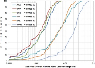
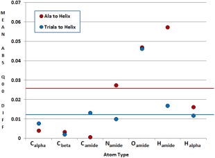
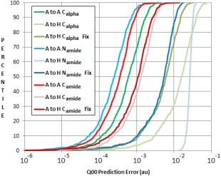
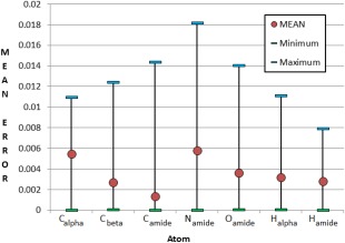
Continuing the development of the FFLUX, a multipolar polarizable force field driven by machine learning, we present a modern approach to atom-typing and building transferable models for predicting atomic properties in proteins. Amino acid atomic charges in a peptide chain respond to the substitution of a neighboring residue and this response can be categorized in a manner similar to atom-typing. Using a machine learning method called kriging, we are able to build predictive models for an atom that is defined, not only by its local environment, but also by its neighboring residues, for a minimal additional computational cost. We found that prediction errors were up to 11 times lower when using a model specific to the correct group of neighboring residues, with a mean prediction of ∼0.0015 au. This finding suggests that atoms in a force field should be defined by more than just their immediate atomic neighbors. When comparing an atom in a single alanine to an analogous atom in a deca-alanine helix, the mean difference in charge is 0.026 au. Meanwhile, the same difference between a trialanine and a deca-alanine helix is only 0.012 au. When compared to deca-alanine models, the transferable models are up to 20 times faster to train, and require significantly less ab initio calculation, providing a practical route to modeling large biological systems. © 2016 The Authors. Journal of Computational Chemistry Published by Wiley Periodicals, Inc.

Keywords: QTAIM; atomic charge; force field design; kriging; machine learning; peptides; quantum chemical topology.


Figures

Figure 1
Figure 1
Superposition of the molecular graph and topological atoms in (top panel) “trialanine” (AAA, 42 atoms) and (bottom panel) the central fragment (10 atoms). The space‐filling nature of the atoms makes it easy to isolate molecular fragments. The hydrogen atom that is bonded to the Cα is hidden in both panels. [Color figure can be viewed at wileyonlinelibrary.com]
Figure 2
Figure 2
Two‐dimensional illustrations of each the three types of peptide data set. Schematic images of molecules are not representative of their actual geometries. Carbon atoms (black) are not labeled and hydrogen atoms are not shown. [Color figure can be viewed at wileyonlinelibrary.com]
Figure 3
Figure 3
A set of 100 geometries for the tripeptide GAG undergoes a series of substitutions (GAG, GAA, AAA, VAA, VAV, see Data Set 2, Fig. 2) and its central alanine's Calpha charges (Q 00, given by the program AIMAll) are plotted as the change between each substitution. For example, the green series gives the difference in charges (obtained through ab initio calculation) between the tripeptides AAA and VAA in Data Set 2. [Color figure can be viewed at wileyonlinelibrary.com]
Figure 4
Figure 4
A set of 100 geometries for the tripeptide GAG undergoes a series of substitutions (GAG, GAA, AAA, VAA, VAV, see Data Set 2 in Fig. 2) and its central alanine's Calpha charges (given by the program AIMAll) are plotted. Each geometry number has a single set of tripeptides (GAG, GAA, AAA, VAA, VAV) that share an identical backbone geometry. The difference in charge between geometries can be seen when comparing the results for different geometry numbers (x‐axis) while the difference in charge due to different terminal sidechains can be seen when comparing tripeptides of the same geometry number. [Color figure can be viewed at wileyonlinelibrary.com]
Figure 5
Figure 5
Comparison of kriging prediction of a central Calpha atom's charge for six tripeptides. Tripeptides AAA, VAV, GAG are part of “group A” (Table 1). Tripeptides YAY, KAK, WAW are part of group D. The kriging model was trained for the tripeptide AAA (trialanine) using geometries that are not present in the test set. Mean prediction errors for the Calpha charges of each tripeptide are given in au. [Color figure can be viewed at wileyonlinelibrary.com]
Figure 6
Figure 6
The difference in atomic charge between atoms in alanine, trialanine and a deca‐alanine helix. A solid line shows the overall mean difference in charges for each series (0.026 au and 0.012 au for alanine and trialanine, respectively). The mean difference across all 2000 geometries is expressed per atom. [Color figure can be viewed at wileyonlinelibrary.com]
Figure 7
Figure 7
Kriging models for the Calpha of a single alanine predicting Calpha charges in alanine (A) and in a deca‐alanine helix (H). “μ Fix” predictions show predictions of helix Calpha properties with altered μ values in the kriging models. [Color figure can be viewed at wileyonlinelibrary.com]
Figure 8
Figure 8
Prediction of charges in a deca‐alanine helix from models trained using trialanine data (AAA to H). All charges are expressed in atomic units. [Color figure can be viewed at wileyonlinelibrary.com]

Similar articles

Cited by

References

    1. Wishart D. S., Bigam C. G., Holm A., Hodges R. S., Sykes B. D., J. Biomol. NMR 1994, 5, 67. - PubMed
    1. Monaco G., Int. J. Quantum Chem. 1998, 68, 201.
    1. Thomas A., Milon A., Brasseur R., Proteins Struct. Funct. Bioinform. 2004, 56, 102. - PubMed
    1. Rypniewski W. R., Perrakis A., Vorgias C. E., Wilson K. S., Protein Eng. 1994, 7, 57. - PubMed
    1. Brooks B. R., Brooks C. L. I., MacKerell A. J., J. Comput. Chem. 2009, 30, 1545. - PMC - PubMed

Publication types

LinkOut - more resources