. 2017 Feb 1;127(2):487-499.
doi: 10.1172/JCI87526. Epub 2016 Dec 19.

M1 muscarinic allosteric modulators slow prion neurodegeneration and restore memory loss

M1 muscarinic allosteric modulators slow prion neurodegeneration and restore memory loss

Sophie J Bradley et al. J Clin Invest. .

Abstract

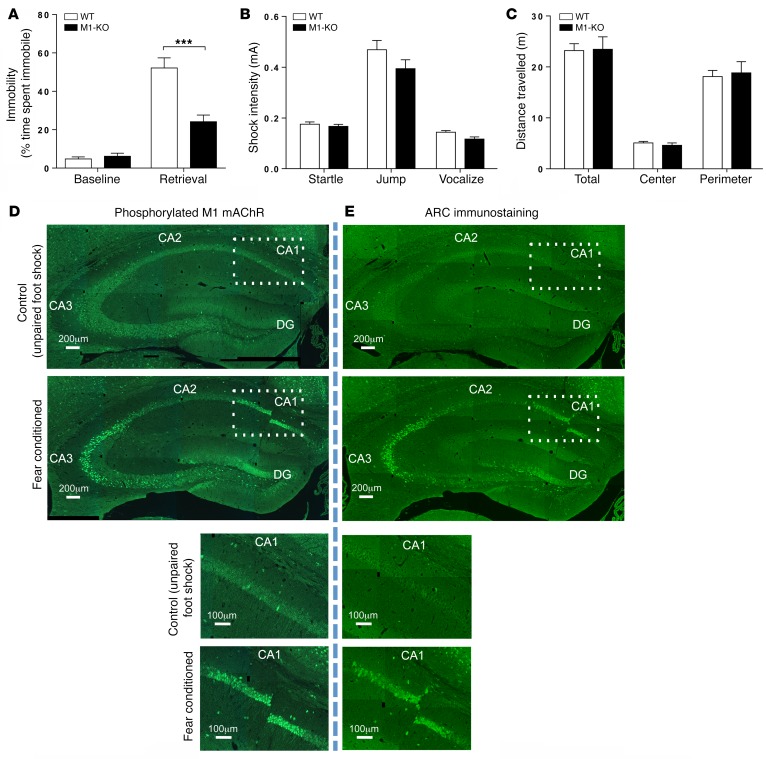
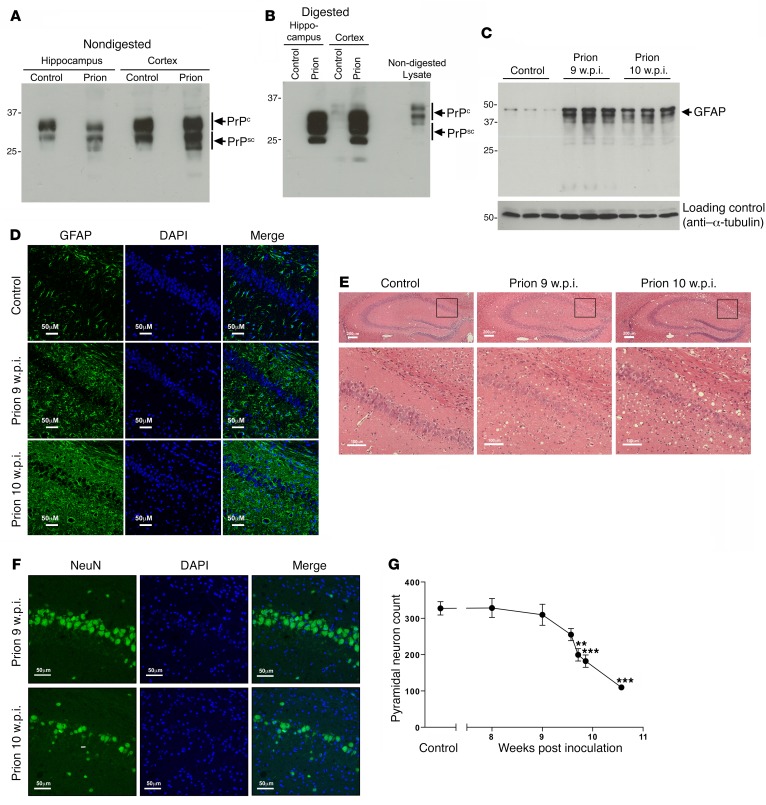
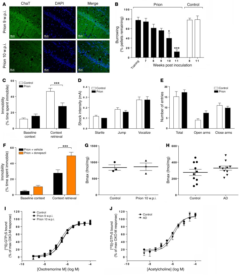
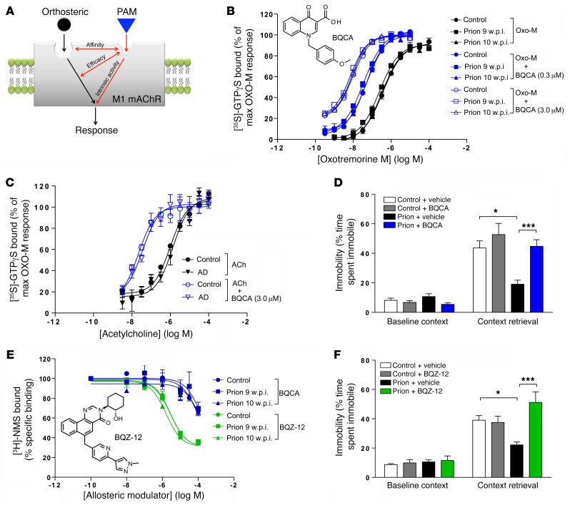
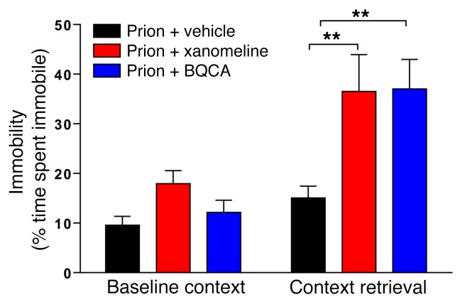
The current frontline symptomatic treatment for Alzheimer's disease (AD) is whole-body upregulation of cholinergic transmission via inhibition of acetylcholinesterase. This approach leads to profound dose-related adverse effects. An alternative strategy is to selectively target muscarinic acetylcholine receptors, particularly the M1 muscarinic acetylcholine receptor (M1 mAChR), which was previously shown to have procognitive activity. However, developing M1 mAChR-selective orthosteric ligands has proven challenging. Here, we have shown that mouse prion disease shows many of the hallmarks of human AD, including progressive terminal neurodegeneration and memory deficits due to a disruption of hippocampal cholinergic innervation. The fact that we also show that muscarinic signaling is maintained in both AD and mouse prion disease points to the latter as an excellent model for testing the efficacy of muscarinic pharmacological entities. The memory deficits we observed in mouse prion disease were completely restored by treatment with benzyl quinolone carboxylic acid (BQCA) and benzoquinazoline-12 (BQZ-12), two highly selective positive allosteric modulators (PAMs) of M1 mAChRs. Furthermore, prolonged exposure to BQCA markedly extended the lifespan of diseased mice. Thus, enhancing hippocampal muscarinic signaling using M1 mAChR PAMs restored memory loss and slowed the progression of mouse prion disease, indicating that this ligand type may have clinical benefit in diseases showing defective cholinergic transmission, such as AD.


Conflict of interest statement

The authors have declared that no conflict of interest exists.

Figures

Figure 1
Figure 1. M1 mAChRs play an important role in hippocampal-dependent learning and memory.
(A) Fear-conditioning response of WT and M1-KO mice. Statistical analysis by 2-way ANOVA with Sidak’s multiple comparison test. ***P < 0.001. (B) Pain thresholds for WT and M1-KO mice. Statistical analysis by Student’s t test. (C) Locomotion of WT and M1-KO mice was determined by total distance traveled during an open field test. Data were analyzed using 2-way ANOVA with Sidak’s multiple comparisons. All WT and M1-KO behavioral data are shown as mean ± SEM of n = 8 mice. (D) An antibody-based biosensor for M1 mAChR activation (phosphorylation of the M1 mAChR on S228 in the third intracellular loop) was used to assess M1 mAChR activity in the hippocampus. Following fear-conditioning training, phosphorylation at S228 of the M1 mAChR was increased in the CA1 and CA3 regions and dentate gyrus of the hippocampus relative to control mice that received a 2-second unpaired foot shock. Magnification of the CA1 region (indicated by the rectangle) is shown in lower panels. (E) Fear-conditioning training increased neuronal activity, as assessed by an increase in ARC immunostaining, in the same regions of the hippocampus as those observed for activated M1 mAChR. D and E are composited images. Magnification of the CA1 region (indicated by the rectangle) is shown in lower panels (see D and E). Scale bars: 200 μm (upper panels); 100 μm (lower panels).
Figure 2
Figure 2. Mouse prion disease is associated with the accumulation of misfolded prion protein, astrogliosis, spongiosis, and hippocampal neuronal loss.
(A) Lysates from control or prion-infected (10 w.p.i.) mouse hippocampus and cortex were probed in Western blots with an antibody that detected both cellular (PrPc) and misfolded (PrPSc) prion protein. The presence of misfolded PrPsc is evident by lower molecular weight species in the prion-infected lysates. (B) Lysates from A were treated with proteinase K before probing in Western blots for prion protein. (C) Astrogliosis during prion disease was determined in Western blots of lysates prepared from control mice or mice 9 and 10 w.p.i. and probed with glial fibrillary acidic protein (GFAP), a marker for astrocytes. (D) Immunohistochemical staining of the hippocampal CA1 region probed with anti–glial fibrillary acidic protein antibody (green) to determine the level of astrogliosis. The nuclei were stained blue with DAPI. Scale bars: 50 μm. (E) Spongiosis in prion-infected hippocampi (upper panels) and CA1 region (lower panels) was visualized by H&E stain of mouse hippocampus from paraformaldehyde-fixed mouse brain from control mice injected with NBH (control), prion-infected mice 9 w.p.i., and prion-infected mice 10 w.p.i. Scale bars: 200 μm (upper panels); 100 μm (lower panels). (F) Determination of neuronal loss of pyramidal neurons in the CA1 region of the hippocampus of mice 9 and 10 w.p.i. was determined by immunohistochemical staining of neuronal cell bodies using antibodies to NeuN (green). The nuclei were stained blue with DAPI. Scale bars: 50 μm. (G) Quantification of NeuN staining in the CA1 of mice at various stages of prion disease. Data are shown as mean ± SEM. n = 3 mice, 3 sections per mouse. **P < 0.01; ***P < 0.001, 1-way ANOVA. Blots and images shown are representative of at least 3 independent experiments.
Figure 3
Figure 3. Prion disease is associated with a disruption in hippocampal cholinergic innervation, a deficit in learning, and memory rescued by donepezil, while maintaining muscarinic receptor expression and signaling.
(A) Cholinergic innervation of the hippocampus was assessed by ChAT (green) immunostaining of the CA1 region of the hippocampus in mice at 9 and 10 w.p.i. Data shown are representative of 3 individual mice per group. Scale bars: 50 μm. (B) Burrowing response of control and prion-infected mice. n = 4–9 mice. *P < 0.05; ***P < 0.001, 1-way ANOVA. (C) Fear-conditioning response of control and prion-infected mice at 9–10 w.p.i. n = 19 mice per group. ***P < 0.001, 2-way ANOVA with Sidak’s multiple comparison test. (D) Pain threshold response of control and prion-infected mice at 9–10 w.p.i. n = 6 mice per group. Unpaired Student’s t test. (E) Anxiety levels of control and prion-infected mice at 9–10 w.p.i. were assessed by elevated plus maze. n = 6 mice per group. Unpaired Student’s t test. (F) Fear-conditioning response of prion-infected mice (9–10 w.p.i.) treated with vehicle or donepezil (0.5 mg/kg) 60 minutes before training. n = 9 (vehicle); n = 15 (donepezil). ***P < 0.001, 2-way ANOVA with Sidak’s multiple comparison test. (G) Determination of the total muscarinic receptor population by [3H]-NMS binding to hippocampal membranes prepared from control or prion-infected mice (10 w.p.i.). Nonspecific binding was determined by the addition of atropine (1 μM). Data are expressed as fmol/mg protein (n = 3). Bmax, maximal binding capacity. (H) Total [3H]-NMS binding to membranes prepared from the frontal cortex of control or AD patients. n = 10. (I) Stimulation of [35S]-GTPγS binding to membranes prepared from control or prion-infected mice (9–10 w.p.i.) in response to oxotremorine-M. Data shown are increases in [35S]-GTPγS binding over basal; mean pEC50 values of 6.45 ± 0.03, 6.63 ± 0.01, and 6.50 ± 0.04, respectively (n = 3). (J) [35S]-GTPγS binding to membranes prepared from the frontal cortex of control or AD patients in response to acetylcholine. Data are the percentage of the maximal [35S]-GTPγS binding stimulated by oxotremorine-M. Mean pEC50 values of 6.00 ± 0.09 and 5.86 ± 0.11, respectively (n = 3).
Figure 4
Figure 4. The orthosteric mAChR agonist xanomeline restores the learning and memory deficit in prion-infected mice.
(A) Chemical structure of xanomeline. (B) [35S]-GTPγS binding to membranes prepared from control or prion-infected mice (9–10 w.p.i.) in response to xanomeline are expressed as a percentage of the maximal response observed with oxotremorine-M. Mean pEC50 for xanomeline on control membranes = 7.67 ± 0.04, prion 9 w.p.i. membranes = 7.73 ± 0.06, and prion 10 w.p.i. membranes = 7.65 ± 0.13. n = 3. (C) [35S]-GTPγS binding to membranes prepared from the frontal cortex of control or AD patients in response to xanomeline. Data are expressed as the percentage of the maximal [35S]-GTPγS binding stimulated by oxotremorine-M. Mean pEC50 values of 8.14 ± 0.28 (control) and 7.34 ± 0.36 (AD). n = 3. (D) Fear-conditioning response of control and prion-infected mice following administration of vehicle or xanomeline (5 mg/kg) 30 minutes prior to training and retrieval. n ≥ 6. Statistical analysis by 1 -way ANOVA. **P < 0.01. (E) Burrowing response of control and prion-infected mice following administration of vehicle or xanomeline (5 mg/kg) 30 minutes before each burrowing session (from 7 w.p.i.). (F, G, and H ) AUC of AMPA receptor–mediated currents before and after treatment with xanomeline in control (F, n = 10) and prion-infected (G and H, n = 12) hippocampi. *P < 0.05, paired Student’s t test. Also shown in G are representative traces of paired whole cell CA1 glutamatergic current recordings in vehicle-treated (black) and xanomeline-treated (100 nM) (red) hippocampal slices of a prion-infected mouse. (I) Western blot of hippocampal lysates prepared from prion-infected mice treated with vehicle or xanomeline (Xan, 5 mg/kg) and probed with an antibody that detects phospho-S880 of GluR2 AMPA receptor subunits (total GluR2 was used as a loading control). (J) Quantification of I. n = 3. **P < 0.01, paired Student’s t test. Data are shown as mean ± SEM.
Figure 5
Figure 5. PAMs of the M1 mAChR rescue the fear-conditioning learning and memory deficit in prion-infected mice.
(A) Schematic summarizing the 3 possible effects of an allosteric modulator, namely, modulation of orthosteric ligand affinity, signaling efficacy, and/or direct activation. (B) BQCA (inset; chemical structure of BQCA) causes equivalent leftward shifts (black arrow) of the oxotremorine-M (Oxo-M) [35S]-GTPγS-assay concentration-response curve and displays intrinsic activity (red arrow) in hippocampal membranes derived from control and prion-infected mice (9 and 10 w.p.i.). Data are shown as mean ± SEM. n = 3. (C) Acetylcholine-stimulated [35S]-GTPγS binding to membranes prepared from the frontal cortex of control or AD patients in the absence and presence of BQCA (3 μM). Data are expressed as the percentage of the maximal [35S]-GTPγS binding stimulated by oxotremorine-M. Mean ± SEM. n = 3. (D) Fear-conditioning response of control and prion-infected mice following administration of vehicle or BQCA (15 mg/kg) 30 minutes prior to training. Mean ± SEM. n = 6–18. *P < 0.05; ***P < 0.001, 1-way ANOVA. (E) Radioligand competition binding between [3H]-NMS (~0.3 nM) and increasing concentrations of BQCA or BQZ-12 (inset, chemical structure of BQZ-12) in hippocampal membranes from control and prion-infected mice (9 and 10 w.p.i.). n = 3–4. The affinities (pKi) of BQCA and BQZ-12 at hippocampal membranes from prion-diseased mice (10 w.p.i.) were 6.15 ± 0.08 and 4.25 ± 0.12, respectively. Data are shown as mean ± SEM. n = 3. (F) Fear-conditioning response of control and prion-infected mice following administration of vehicle or BQZ-12 (1.5 mg/kg) 30 minutes prior to training. Data are shown as mean ± SEM. n = 12–19. *P < 0.05; ***P < 0.001, 1-way ANOVA.
Figure 6
Figure 6. Orthosteric agonists and PAMs of the M1 mAChR are equivalently efficacious in restoring fear-conditioning learning and memory deficit in prion-infected mice.
Fear-conditioning response of prion-infected mice following administration of vehicle, xanomeline (5 mg/kg), or BQCA (15 mg/kg) 30 minutes prior to training (n = 13–18). Data are shown as mean ± SEM. **P < 0.01, 1-way ANOVA.
Figure 7
Figure 7. PAMs of the M1 mAChR significantly increase survival in prion-diseased mice.
Kaplan-Meier survival plots for prion-infected mice treated with vehicle (5% glucose; n = 10; black line) or BQCA (15 mg/kg; n = 10; blue line) daily from 7 w.p.i. ***P < 0.001, Gehan-Breslow-Wilcoxon test.

References

    1. Altenbach C, Kusnetzow AK, Ernst OP, Hofmann KP, Hubbell WL. High-resolution distance mapping in rhodopsin reveals the pattern of helix movement due to activation. Proc Natl Acad Sci U S A. 2008;105(21):7439–7444. doi: 10.1073/pnas.0802515105. - DOI - PMC - PubMed
    1. Massoud F, Gauthier S. Update on the pharmacological treatment of Alzheimer’s disease. Curr Neuropharmacol. 2010;8(1):69–80. doi: 10.2174/157015910790909520. - DOI - PMC - PubMed
    1. Coyle JT, Price DL, DeLong MR. Alzheimer’s disease: a disorder of cortical cholinergic innervation. Science. 1983;219(4589):1184–1190. doi: 10.1126/science.6338589. - DOI - PubMed
    1. LaFerla FM, Green KN, Oddo S. Intracellular amyloid-beta in Alzheimer’s disease. Nat Rev Neurosci. 2007;8(7):499–509. doi: 10.1038/nrn2168. - DOI - PubMed
    1. Nathan PJ, et al. The potent M1 receptor allosteric agonist GSK1034702 improves episodic memory in humans in the nicotine abstinence model of cognitive dysfunction. Int J Neuropsychopharmacol. 2013;16(4):721–731. doi: 10.1017/S1461145712000752. - DOI - PubMed

MeSH terms

Substances