. 2017 Feb;24(2):131-139.
doi: 10.1038/nsmb.3344. Epub 2016 Dec 19.

Structural basis for targeted DNA cytosine deamination and mutagenesis by APOBEC3A and APOBEC3B

Affiliations

Structural basis for targeted DNA cytosine deamination and mutagenesis by APOBEC3A and APOBEC3B

Ke Shi et al. Nat Struct Mol Biol. 2017 Feb.

Abstract

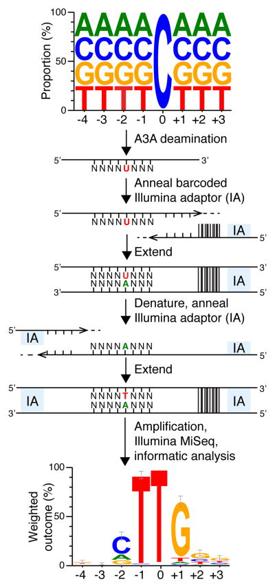
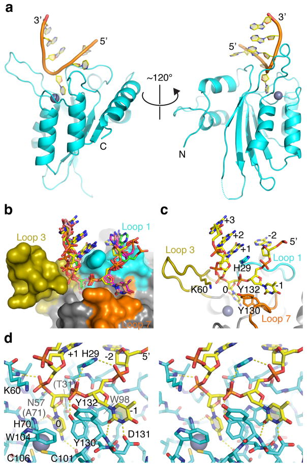
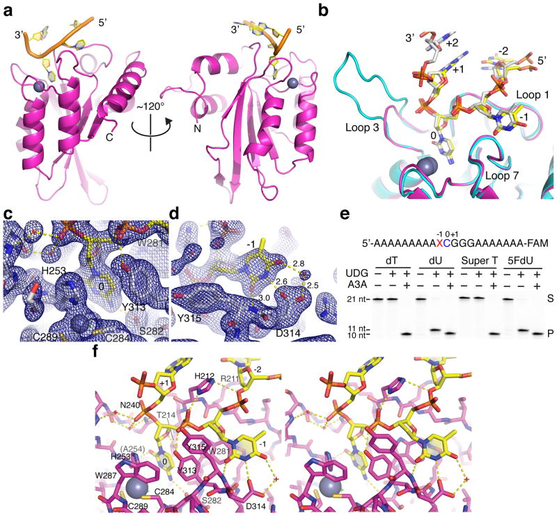
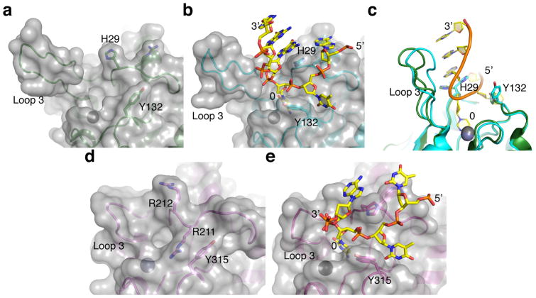
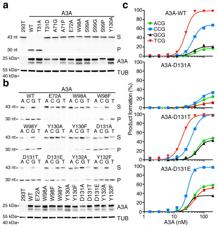
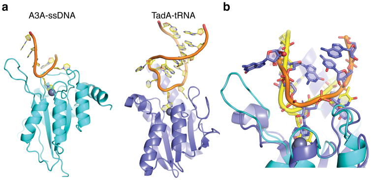
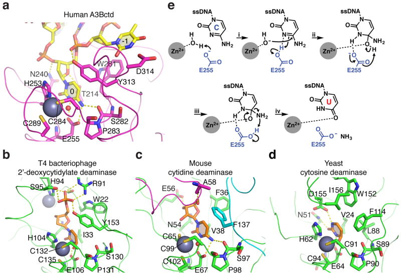
APOBEC-catalyzed cytosine-to-uracil deamination of single-stranded DNA (ssDNA) has beneficial functions in immunity and detrimental effects in cancer. APOBEC enzymes have intrinsic dinucleotide specificities that impart hallmark mutation signatures. Although numerous structures have been solved, mechanisms for global ssDNA recognition and local target-sequence selection remain unclear. Here we report crystal structures of human APOBEC3A and a chimera of human APOBEC3B and APOBEC3A bound to ssDNA at 3.1-Å and 1.7-Å resolution, respectively. These structures reveal a U-shaped DNA conformation, with the specificity-conferring -1 thymine flipped out and the target cytosine inserted deep into the zinc-coordinating active site pocket. The -1 thymine base fits into a groove between flexible loops and makes direct hydrogen bonds with the protein, accounting for the strong 5'-TC preference. These findings explain both conserved and unique properties among APOBEC family members, and they provide a basis for the rational design of inhibitors to impede the evolvability of viruses and tumors.


Conflict of interest statement

RSH and DAH are co-founders, shareholders, and consultants of ApoGen Biotechnologies Inc. HA and REA are consultants for ApoGen Biotechnologies Inc. REA is a co-founder of Actavalon Inc. The other authors have no competing financial interests to declare.

Figures

Figure 1
Figure 1. Deep-deamination approach to determine an optimal human A3A substrate
A ssDNA library with a single target C and N’s on the 5′ and 3′ sides was reacted with human A3A (near-single hit kinetics). The resulting pool containing C-to-U deamination products was annealed to a bar-coded Illumina adaptor (IA) and T4 DNA polymerase was used to produce a complementary DNA strand. This intermediate was denatured, annealed to a 5′-IA, and converted to duplex by Phusion thermostable high fidelity DNA polymerase. Illumina Mi-Seq was used to generate reads for subsequent informatics analysis. A weblogo representation of deamination products unique to A3A shows enrichment for −1 T and +1 G that informed the ssDNA sequence for co-crystallization experiments (n=641; error bars are twice the sample correction value). Source data are provided as a spreadsheet (.xlsx) linked to the online legend.
Figure 2
Figure 2. Crystal structure of human A3A bound to ssDNA with preferred 5′-TCG deamination target motif
(a) Ribbon schematic of A3A-ssDNA complex showing flipped-out target C and −1 T nucleotides, as well as the overall U-shaped binding conformation. (b) Molecular surface of A3A active site with the surrounding loops color-coded, and a superposition of stick-models of ssDNA bound to 4 different molecules in the crystal’s asymmetric unit. (c) A view similar to panel b with a representative ssDNA molecule shown and nucleobases and key amino side chains from the active site loops labeled. (d) A wall-eye stereo view of the A3A active site and the bound ssDNA molecule shown in sticks. Hydrogen bonds are indicated by yellow dashed lines.
Figure 3
Figure 3. Crystal structure of a variant of the human A3B catalytic domain bound to ssDNA with a 5′-TCA deamination target motif
(a) Ribbon schematic of ssDNA-A3Bctd* complex showing the flipped-out target C (0) and −1 T, as well as the overall U-shaped binding conformation. (b) A superposition of the active site region of A3A (cyan) and A3Bctd* (magenta) with relevant ssDNA substrates (opaque from a representative A3A structure and yellow from the A3Bctd* structure) showing the near-identical positioning of the flipped-out target C and −1 T. (c–d) Composite omit 2Fo-Fc map contoured at 1.0σ shown for region surrounding the target cytosine (panel c) or −1 thymine (panel d). (e) Deaminase activity of wild-type A3A on ssDNA substrates containing normal T or the indicated analogs at the −1 position demonstrating that the 5-methyl group is unconstrained structurally. Uracil DNA glycosylase (UDG) readily excises dU and 5FdU from ssDNA and accounts for the 11-nucleotide product in the absence of deamination. However, due to A3A activity on the target C and the 3′-end label, only the shorter 10-nucleotide product is apparent upon deamination and gel fractionation. The uncropped gel image is provided in Supplementary Data Set 1, and the results are representative of 2 independent experiments. (f) A wall-eye stereo view of the A3Bctd* active site and the bound ssDNA molecule shown in sticks. Hydrogen bonds are indicated by yellow dashed lines. Water molecules are represented by small red crosshairs.
Figure 4
Figure 4. Comparison between apo and ssDNA-bound A3A and A3B structures
(a–b) Molecular surface of A3A around the active site in the DNA-free (panel a, pdb 4XXO) and ssDNA-bound (panel b, this study) states, showing re-orientation of the side chains of His29 and Tyr132. (c) A superposition of the two conformations in panels a and b highlighting the repositioning of His29 and Tyr132 as well as shifting of loop 3 toward the bound ssDNA. (d–e) Molecular surface of A3Bctd* around the active site in the DNA-free (panel d, pdb 5CQH) and ssDNA-bound (panel e, this study) states showing the large transition that is likely to occur between the closed (unbound) and open (ssDNA-bound) conformations.
Figure 5
Figure 5. Corroborating biochemical data for human A3A
(a–b) DNA cytosine deamination by human A3A (WT, wild-type) and the indicated mutant derivatives (S, substrate; P, product). The corresponding anti-MYC (A3A) and anti-TUBULIN immunoblots indicate similar levels of A3A and soluble extract in each experiment relative to controls (A3A has two bands due to alternative translation initiation from Met1 or Met13). Reactions in panel a interrogated active site mutants using a 43-nucleotide 5′-TC-containing ssDNA substrate, and reactions in panel b additionally interrogated the identity of the −1 position (A, C, G, or T) relative to the target cytosine. These results are representative of 2 independent experiments, and uncropped gel images are provided in Supplementary Data Set 1. (c) Graphs quantifying product accumulation in dose response experiments for A3A and the indicated D131 derivatives using the same ssDNA substrates as panel b. WT enzyme and D131T show similar activities and local −1 nucleobase preferences, D131A has low activity and relaxed preferences, and D131E has slightly lower activity and a clear preference for −1 C instead of −1 T. These data are representative of 2 independent experiments, and source data are provided as a spreadsheet (.xlsx) linked to the online legend.
Figure 6
Figure 6. Human A3A and S. aureus TadA have similar U-shaped polynucleotide binding conformations
(a) Ribbon schematics of A3A-ssDNA (this study) and TadA-tRNA (pdb 2B3J) with the single zinc-coordinating active site regions positioned at similar angles for comparison. (b) Superposition of A3A-ssDNA and TadA-tRNA structures showing similar U-shaped binding conformations (predicted in a commentary to the original TadA-tRNA structure report). A3A-bound ssDNA is shown in yellow and TadA-bound RNA is shown with an orange backbone and magenta nucleobases.
Figure 7
Figure 7. Structural comparison of the active sites of A3B and distantly related deaminase family members
Active sites of (a) A3Bctd bound to ssDNA, (b) T4 bacteriophage 2′-deoxycytidylate deaminase bound to a dCMP analog (pdb 1VQ2), (c) murine cytidine deaminase bound to cytidine (pdb 2FR6), and (d) yeast cytosine deaminase bound to an analog of the free nucleobase cytosine (pdb 1P6O). The catalytic glutamate (Glu255) was modeled into the A3Bctd*-ssDNA structure based on its positioning in the apo structure (pdb 5CQH), closely mimicking conformations of the corresponding residues in the T4, mouse, and yeast enzymes (Glu106, Glu67, and Glu64, respectively). Gray spheres represent zinc ions. Smaller red spheres show the zinc-bound reactive water molecule. Sticks depict key residues contacting bound substrates, and ribbons represent protein backbones. The magenta loop in panel c is from an adjacent subunit in the tetramer. A comparison of the structures shows similar zinc-coordination mechanisms and target cytosine positioning including conservation of surrounding aromatic residues, as well as substrate-specific interactions conferred by unique residues for each class of enzyme. (e) Proposed deamination mechanism (adapted from ref.): (i) Glu255 deprotonates the Zn2+-coordinated H2O for nucleophilic attack at cytosine with the residual, protonated Glu255 H-bonding to N3 to withdraw electron density from C4 of cytosine, thereby accelerating nucleophilic attack by hydroxide. (ii) Deprotonation of the alcohol on the tetrahedral intermediate by Glu255 ensues, followed by (iii) collapse of the tetrahedral intermediates due to elimination of ammonia, to which protonated Glu255 contributes a hydrogen. (iv) The resulting uracil base is likely stabilized by Zn2+ coordination in the enzyme active site.

References

    1. Conticello SG. The AID/APOBEC family of nucleic acid mutators. Genome Biol. 2008;9:229. - PMC - PubMed
    1. Harris RS, Dudley JP. APOBECs and virus restriction. Virology. 2015;479–480C:131–145. - PMC - PubMed
    1. Di Noia JM, Neuberger MS. Molecular mechanisms of antibody somatic hypermutation. Annu Rev Biochem. 2007;76:1–22. - PubMed
    1. Robbiani DF, Nussenzweig MC. Chromosome translocation, B cell lymphoma, and activation-induced cytidine deaminase. Annu Rev Pathol. 2013;8:79–103. - PubMed
    1. Fossat N, Tam PP. Re-editing the paradigm of cytidine (C) to uridine (U) RNA editing. RNA Biol. 2014;11:1233–1237. - PMC - PubMed

Publication types

MeSH terms