

Posttraumatic Stress Disorder: Perspectives for the Use of Deep Brain Stimulation
- PMID: 27992092
- PMCID: PMC5247323
- DOI: 10.1111/ner.12551
Posttraumatic Stress Disorder: Perspectives for the Use of Deep Brain Stimulation
Abstract
Objectives: Deep Brain Stimulation (DBS) has been either approved or is currently under investigation for a number of psychiatric disorders.
Materials and methods: We review clinical and preclinical concepts as well as the neurocircuitry that may be of relevance for the implementation of DBS in posttraumatic stress disorder (PTSD).
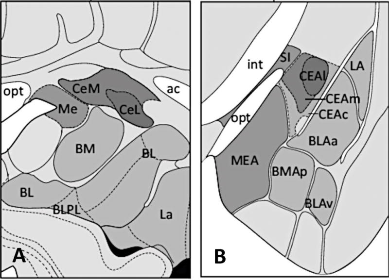
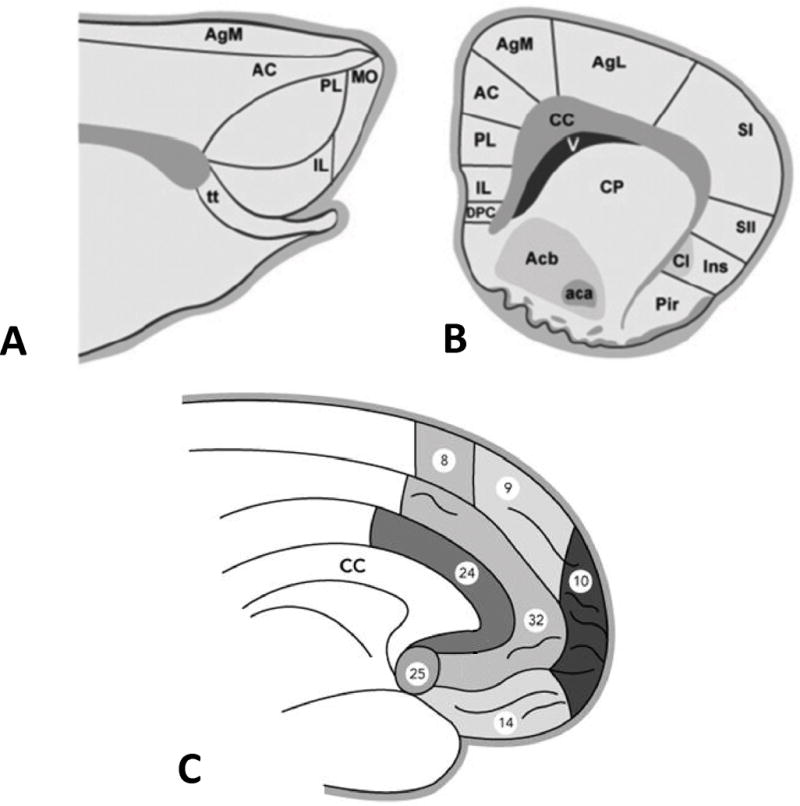
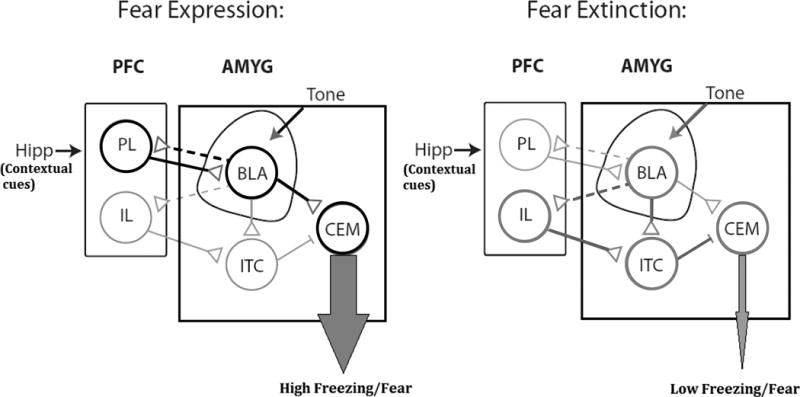
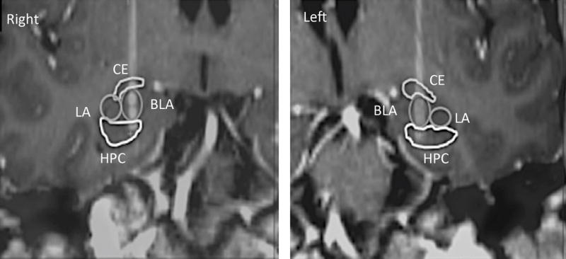
Results: PTSD is a chronic and debilitating illness associated with dysfunction in well-established neural circuits, including the amygdala and prefrontal cortex. Although most patients often improve with medications and/or psychotherapy, approximately 20-30% are considered to be refractory to conventional treatments. In other psychiatric disorders, DBS has been investigated in treatment-refractory patients. To date, preclinical work suggests that stimulation at high frequency delivered at particular timeframes to different targets, including the amygdala, ventral striatum, hippocampus, and prefrontal cortex may improve fear extinction and anxiety-like behavior in rodents. In the only clinical report published so far, a patient implanted with electrodes in the amygdala has shown striking improvements in PTSD symptoms.
Conclusions: Neuroimaging, preclinical, and preliminary clinical data suggest that the use of DBS for the treatment of PTSD may be practical but the field requires further investigation.
Keywords: Amygdala; anxiety; deep brain stimulation; fear extinction; posttraumatic stress disorder; prefrontal cortex.
© 2016 International Neuromodulation Society.
Conflict of interest statement
The authors have no conflicts of interest to declare.
Figures
References
-
- American Psychiatric Association. Diagnostic and statistical manual of mental disorders (DSM V) 4th. Washington, DC: 2013. Revised.
-
- Kessler RC, Berglund P, Demler O, Jin R, Merikangas KR, Walters EE. Lifetime prevalence and age-of-onset distributions of DSM-IV disorders in the National Comorbidity Survey Replication. Arch Gen Psychiatry. 2005;62:593–602. - PubMed
-
- Kessler RC, Sonnega A, Bromet E, Hughes M, Nelson CB. Posttraumatic stress disorder in the National Comorbidity Survey. Arch Gen Psychiatry. 1995;52:1048–1060. - PubMed
-
- Hoge CW, Castro CA, Messer SC, McGurk D, Cotting DI, Koffman RL. Combat duty in Iraq and Afghanistan, mental health problems, and barriers to care. N Engl J Med. 2004;351:13–22. - PubMed
-
- Kang HK, Natelson BH, Mahan CM, Lee KY, Murphy FM. Post-traumatic stress disorder and chronic fatigue syndrome-like illness among Gulf War veterans: a population-based survey of 30,000 veterans. Am J Epidemiol. 2003;157:141–148. - PubMed
Publication types
MeSH terms
Grants and funding
LinkOut - more resources
Full Text Sources
Other Literature Sources
Medical
