. 2017 Feb;49(2):193-203.
doi: 10.1038/ng.3741. Epub 2016 Dec 19.

A genome-wide CRISPR screen identifies a restricted set of HIV host dependency factors

Affiliations

A genome-wide CRISPR screen identifies a restricted set of HIV host dependency factors

Ryan J Park et al. Nat Genet. 2017 Feb.

Abstract

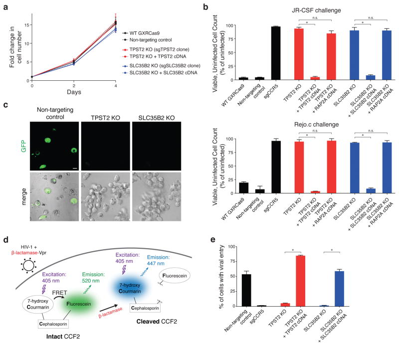
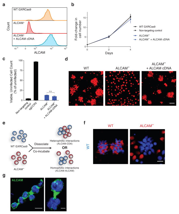
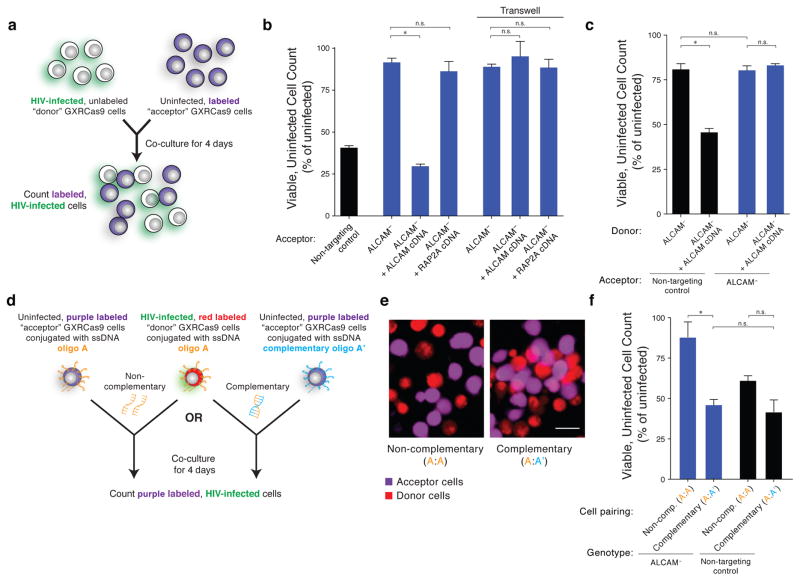
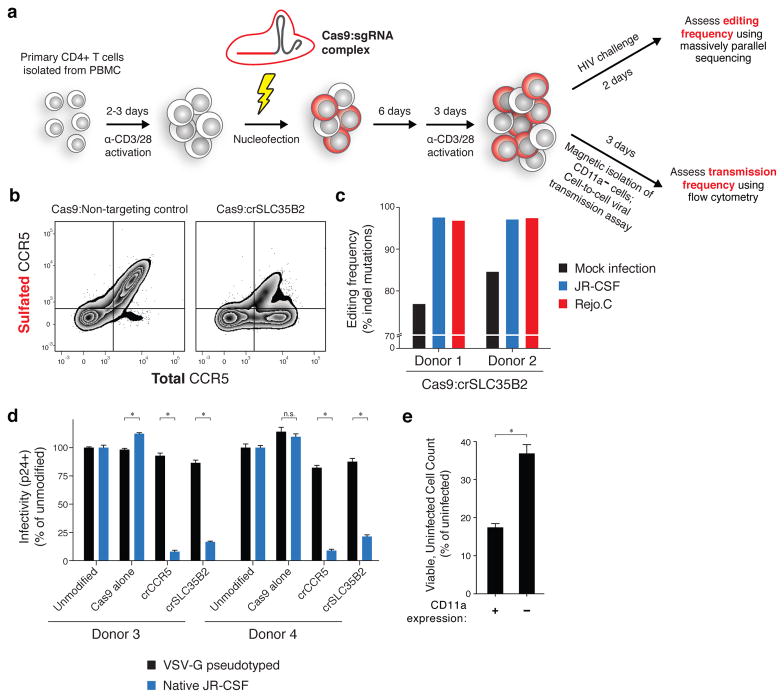
Host proteins are essential for HIV entry and replication and can be important nonviral therapeutic targets. Large-scale RNA interference (RNAi)-based screens have identified nearly a thousand candidate host factors, but there is little agreement among studies and few factors have been validated. Here we demonstrate that a genome-wide CRISPR-based screen identifies host factors in a physiologically relevant cell system. We identify five factors, including the HIV co-receptors CD4 and CCR5, that are required for HIV infection yet are dispensable for cellular proliferation and viability. Tyrosylprotein sulfotransferase 2 (TPST2) and solute carrier family 35 member B2 (SLC35B2) function in a common pathway to sulfate CCR5 on extracellular tyrosine residues, facilitating CCR5 recognition by the HIV envelope. Activated leukocyte cell adhesion molecule (ALCAM) mediates cell aggregation, which is required for cell-to-cell HIV transmission. We validated these pathways in primary human CD4+ T cells through Cas9-mediated knockout and antibody blockade. Our findings indicate that HIV infection and replication rely on a limited set of host-dispensable genes and suggest that these pathways can be studied for therapeutic intervention.


Conflict of interest statement

COMPETING FINANCIAL INTERESTS

T.W., D.M.S., and E.S.L. are inventors on a patent application for functional genomics using the CRISPR-Cas system (US 15/141,348), T.W and D.M.S are founders of KSQ Therapeutics, a CRISPR functional genomics company, and D.M.S is a scientific advisor for KSQ Therapeutics. A patent has been filed on the use of Cas9 RNPs to edit the genome of human primary T cells (A.M. and K.S.). A.M. serves as an advisor to Juno Therapeutics and the Marson lab has had sponsored research agreements with Juno Therapeutics and Epinomics.

Figures

Figure 1
Figure 1. A pooled, genome-wide CRISPR screen for HIV host dependency factors (HDFs)
(a) Outline of genome-wide CRISPR screen strategy. (b) Flow cytometry of cells infected with the HIV-1 strain JR-CSF and expressing GFP as a reporter of productive HIV infection. Where indicated, cells are transduced with sgCCR5 or an sgRNA that does not target protein-coding sequences in the human genome (non-targeting control). (c) Flow cytometry of CD4 and CCR5 surface expression on WT GXRCas9 cells and GXRCas9 cells transduced with the genome-wide sgRNA library and serially infected with the HIV-1 strain JR-CSF. (d) Log2-fold change in abundance of the 5th most enriched sgRNA for every gene following HIV infection. See also Supplementary Table 1 and Supplementary Fig. 1. (e) Enrichment of individual sgRNAs for three candidate HDFs and two control genes. Values indicate log2-fold change in abundance following HIV infection. Uninfected values are from GXRCas9 cells transduced with the genome-wide sgRNA library and cultured for 3 weeks.
Figure 2
Figure 2. Loss of TPST2 or SLC35B2 confers strong protection against HIV infection and entry without compromising host cell viability
(a) Normal proliferation of TPST2-null and SLC35B2-null GXRCas9 cells compared to wild-type cells, non-targeting sgRNA transduced cells, and cells with rescued TPST2 and SLC35B2 expression. Error bars represent standard deviation of six replicate wells. (b) Virus challenge assay with JR-CSF (upper panel) and Rejo.C, a patient-derived transmitted/founder strain of HIV (lower panel). Three days following HIV infection (MOI = 1), viable, GFP-negative cells were counted and normalized to a mock-infected condition. Error bars represent standard deviation of triplicate wells. Error bars, s.d.; triplicate wells; * denotes P<0.01, Welch’s t-test. P-values as follows: JR-CSF – TPST2: *, P<0.0001 and ns, P=0.0637; SLC35B2: *, P=0.0009 and ns, P=0.9714; Rejo.c – TPST2: *, P=0.0005 and ns, P=0.6478; SLC35B2: *, P<0.0001 and ns, P=0.7751. (c) Confocal microscopy of non-targeting control, TPST2-null, and SLC35B2-null GXRCas9 cells following HIV challenge. GFP is a reporter for productive HIV infection. Scale bar = 5 μm. (d) HIV entry assay schematic. β-lactamase-Vpr fusion protein is packaged in HIV virions. Target cells are loaded with CCF2, a FRET donor/acceptor pair linked by a β-lactam ring. Upon viral fusion, the virus-delivered β-lactamase cleaves off the intracellular FRET acceptor, leading to an emission shift. (e) HIV entry assay for TPST2-null and SLC35B2-null GXRCas9 cells compared to wild-type cells, non-targeting control cells, and cells with rescued TPST2 and SLC35B2 expression. Error bars, s.d.; triplicate wells; * denotes P<0.01, Welch’s t-test. P-values as follows: TPST2: *, P<0.0001; SLC35B2: *, P=0.0001.
Figure 3
Figure 3. SLC35B2 and TPST2 act in a common pathway for sulfation of CCR5
(a) HIV entry assay for GXRCas9 cells cultured in standard media or in sulfate-free media with sodium chlorate, an inhibitor of cellular sulfation. Where indicated, heparinase is used to remove cell surface heparan sulfates. Error bars, s.d.; triplicate wells; * denotes P<0.01, Welch’s t-test. P-values as follows: -Heparinase: *, P=0.0005; +Heparinase: *, P<0.0001. (b) Schematic of SLC35B2 and TPST2 sulfating CCR5 within the Golgi apparatus. (c) Flow cytometry of total and sulfated CCR5 surface expression in non-targeting control and CCR5-null GXRCas9 cells, and cells with ablated or rescued TPST2 and SLC35B2 expression.
Figure 4
Figure 4. Homophilic ALCAM interactions are necessary for GXRCas9 cell aggregation
(a) Flow cytometry of surface ALCAM expression in wild-type GXRCas9 cells and cells with ablated or rescued ALCAM expression. (b) Normal proliferation of ALCAM-null cells compared to wild-type, non-targeting control, and ALCAM-rescued cells. Error bars represent standard deviation of six replicate wells. (c) Virus challenge assay with JR-CSF demonstrating that ALCAM-null cells lack protection against HIV infection at an MOI of 1. Error bars, s.d.; triplicate wells; n.s., P=0.0308, Welch’s t-test. (d) Confocal microscopy depicting the cellular aggregation phenotype of GXRCas9 cells with wild-type, null, or rescued ALCAM expression. Scale bar = 50 μm. (e,f) Co-culture assay to distinguish between two models for ALCAM-mediated cell aggregation. (e) Assay design with possible outcomes. Mixed aggregates will result from the co-culture of wild-type and ALCAM-null cells if aggregates are due to heterophilic ALCAM-CD6 interactions while wild-type-only aggregates will result if aggregates are due to homophilic ALCAM-ALCAM interactions. (f) Confocal microscopy demonstrating that ALCAM-null cells are not contained within aggregates. Scale bar = 20 μm. (g) Confocal microscopy of wild-type GXRCas9 cell aggregates for ALCAM (green) and DAPI (blue). Scale bar = 10 μm (left) and 3 μm (right).
Figure 5
Figure 5. Loss of ALCAM protects against cell-to-cell transmission of HIV through disruption of cell aggregation. Error bars represent standard deviation of triplicate wells
(a–c) Cell-to-cell HIV transmission assay. (a) Schematic. Acceptor cells are fluorescently labeled and co-cultured with unlabeled, infected, wild-type donor cells. After 4 days, the number of labeled, GFP-negative, viable cells is assessed by flow cytometry and cell counting and normalized to a matching condition where donor cells are not infected. (b) Donor cells are wild-type and acceptor cells are as indicated on x-axis labels. Where indicated, a 0.45 μm pore size transwell is used to separate donor and acceptor cells for the entirety of the 4 day co-culture. Error bars, s.d.; triplicate wells; *, P<0.0001, Welch’s t-test. n.s. as follows: B vs D, P=0.2605; E vs F, P=0.3481; E vs G, P=0.8903. (c) Donor cells are either null or rescued for ALCAM, and acceptor cells are either wild-type (non-targeting control) or null for ALCAM, as indicated. Error bars, s.d.; triplicate wells; *, P=0.0002, Welch’s t-test. n.s. as follows: A vs C, P=0.8425; C vs D, P=0.1959. (d–f) ALCAM-independent cellular aggregation. (d) Schematic. As with the cell-to-cell HIV transmission assay, donor cells are infected and co-cultured with labeled acceptor cells. Donor and acceptor cells are both ALCAM-null and labeled either with complementary oligonucleotides (A and A′) to induce aggregation, or with identical oligonucleotides (A and A) as a control. (e) Confocal microscopy of oligonucleotide-conjugated cells. Scale bar = 20 μm. (f) Results. Error bars, s.d.; triplicate wells; *, p=0.0092, Welch’s t-test. n.s. as follows: B vs D, P=0.4479; C vs D, P=0.0334.
Figure 6
Figure 6. CRISPR-based approach for validation of HIV host dependency factors in primary human CD4+ T cells
(a) Schematic. Primary CD4+ T cells are activated using antibodies against CD3 and CD28 and nucleofected with Cas9-ribonucleoproteins. After 6 days, cells are re-activated for 3 days and either challenged directly with HIV and sequenced at the sgRNA target site to assess mutation frequency, or purified for CD11a- cells using magnetic beads and used in a cell-to-cell HIV transmission assay. (b) Flow cytometry of total and sulfated CCR5 surface expression in primary CD4+ T cells transfected with Cas9-RNP complexes. Cells were analyzed at the time of HIV infection. (c) Insertion/deletion (indel) mutation frequency in primary CD4+ T cells from two donors following challenge with JR-CSF or Rejo.C, compared to a mock infection condition. See also Supplementary Fig. 4. (d) Flow cytometry of intracellular HIV Gag in Cas9-RNP transfected primary CD4+ T cells from two additional donors. Values are normalized to un-transfected cells for each respective virus type. Entry of VSV-G pseudotyped HIV is independent of CD4 and CCR5. Error bars, s.d.; triplicate wells; * denotes P<0.01, Welch’s t-test. All * P<0.0001 except Donor 3 SLC35B2, P=0.0002. n.s., p=0.1715. (e) Cell-to-cell HIV transmission assay in primary CD4+ T cells transfected with Cas9:crITGAL. Assay is set up as in Figure 5 except donor cells are infected 24 hours prior to co-culture and co-culture is for 2 days. Readout is by flow cytometry following intracellular staining for HIV Gag. Error bars, s.d.; triplicate wells; P=0.0036, Welch’s t-test. See also Supplementary Fig. 6.

Comment in

References

    1. Friedrich BM, Dziuba N, Li G, Endsley MA, et al. Host factors mediating HIV-1 replication. Virus Res. 2011;161:101–114. - PubMed
    1. Fätkenheuer G, Pozniak AL, Johnson MA, Plettenberg A, et al. Efficacy of short-term monotherapy with maraviroc, a new CCR5 antagonist, in patients infected with HIV-1. Nat Med. 2005;11:1170–1172. - PubMed
    1. Gulick RM, Lalezari J, Goodrich J, Clumeck N, et al. Maraviroc for previously treated patients with R5 HIV-1 infection. N Engl J Med. 2008;359:1429–1441. - PMC - PubMed
    1. Tebas P, Stein D, Tang WW, Frank I, et al. Gene editing of CCR5 in autologous CD4 T cells of persons infected with HIV. N Engl J Med. 2014;370:901–910. - PMC - PubMed
    1. Hütter G, Nowak D, Mossner M, Ganepola S, et al. Long-term control of HIV by CCR5 Delta32/Delta32 stem-cell transplantation. N Engl J Med. 2009;360:692–698. - PubMed

MeSH terms