. 2016 Dec 16;11(12):e0166849.
doi: 10.1371/journal.pone.0166849. eCollection 2016.

Simultaneous Administration of ADSCs-Based Therapy and Gene Therapy Using Ad-huPA Reduces Experimental Liver Fibrosis

Affiliations

Simultaneous Administration of ADSCs-Based Therapy and Gene Therapy Using Ad-huPA Reduces Experimental Liver Fibrosis

Alejandra Meza-Ríos et al. PLoS One. .

Abstract

Background and aims: hADSCs transplantation in cirrhosis models improves liver function and reduces fibrosis. In addition, Ad-huPA gene therapy diminished fibrosis and increased hepatocyte regeneration. In this study, we evaluate the combination of these therapies in an advanced liver fibrosis experimental model.

Methods: hADSCs were expanded and characterized before transplantation. Ad-huPA was simultaneously administrated via the ileac vein. Animals were immunosuppressed by CsA 24 h before treatment and until sacrifice at 10 days post-treatment. huPA liver expression and hADSCs biodistribution were evaluated, as well as the percentage of fibrotic tissue, hepatic mRNA levels of Col-αI, TGF-β1, CTGF, α-SMA, PAI-I, MMP2 and serum levels of ALT, AST and albumin.

Results: hADSCs homed mainly in liver, whereas huPA expression was similar in Ad-huPA and hADSCs/Ad-huPA groups. hADSCs, Ad-huPA and hADSCs/Ad-huPA treatment improves albumin levels, reduces liver fibrosis and diminishes Collagen α1, CTGF and α-SMA mRNA liver levels. ALT and AST serum levels showed a significant decrease exclusively in the hADSCs group.

Conclusions: These results showed that combinatorial effect of cell and gene-therapy does not improve the antifibrogenic effects of individual treatments, whereas hADSCs transplantation seems to reduce liver fibrosis in a greater proportion.


Conflict of interest statement

The authors declare that the funder Innovare provided support in the form of salaries for author JAB, but did not have any additional role in the study design, data collection and analysis, decision to publish, or preparation of the manuscript. Besides, author JAB has the USA patent applications numbers 7807457, 7858368 and 8043955 related to application of human uPA wild type/or its truncated version.

Figures

Fig 1
Fig 1. Characterization of hADSCs.
(A) Phenotypic characterization of ADSCs showed cell markers CD105, CD73 and HLA-ABC positive in the population. CD45, CD34 and HLA-DR resulted negative in selected population. (B)Functional characterization of ADSCs into adipogenic, ostegenic and hepatic linage. Photographs (40X) showed non-differentiated cells (control) used as controls of staining and IF and cells positive for lipid droplets (oil red), extracellular calcium deposition (Von Kossa) and IF for AFP and Albumin (FITC).
Fig 2
Fig 2. Biodistribution of hADSCs in cirrhotic rats.
(A) DAPI-stained cells transplanted via iliac vein were detected mainly in liver, and in a very low frequency in lung and spleen. (B) Quantification of DAPI-positive cells per field of view in collected organs (***P<0.001). (C) huPA protein levels in liver homogenates of cirrhotic rats treated with Ad-huPA or the combination of hADSCs and Ad-huPA. Data represent the median for each group.
Fig 3
Fig 3. Assessment of liver fibrosis, inflammation, α-SMA and collagen staining in cirrhotic animals treated with cell and gene therapy.
(A) Macroscopic appearance of the liver and Masson staining showed characteristic morphological liver alterations in cirrhotic rats. Animals treated with any of the therapies showed reduction of fibrotic tissue and decrease in inflammatory cell infiltrate in H&E-stained liver. IHC for α-SMA revealed decrease in staining in treated animals. Sirius red staining indicates a reduction in liver collagen content in animals that received treatment. (B) Fibrosis percentage in liver tissue was measured in control and treated rats (C) Quantification of α-SMA immunoreactivity in liver tissue of treated animals and controls. Data represent the median for each group (*P<0.05, **P<0.01, ***P<0.001). (D) Morphometric quantification of collagen staining using Sirius Red in liver tissue. Data represent the median for each group (*P<0.05, **P<0.01, ***P<0.001).
Fig 4
Fig 4. Analysis of profibrogenic gene expression.
Mean relative expression levels of α-SMA, Col-α1, CTGF, MMP2, PAI-I and TGF-β1 in liver tissue. Data are normalized to GAPDH expression levels and represent the median for each group (*P<0.05, **P<0.01, ***P<0.001).
Fig 5
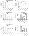
Fig 5. Serum levels of hepatic enzymes.
(A) Liver synthesized albumin augmented in groups that receive cell or/and gene therapy compared to cirrhotic group. (B,C)Transaminases (ALT and AST) showed a diminution only in animals treated with hADSCs. Data represent the median for each group (*P<0.05, **P<0.01, ***P<0.001).

References

    1. Lin H, Xu R, Zhang Z, Chen L, Shi M, Wang FS. Implications of the immunoregulatory functions of mesenchymal stem cells in the treatment of human liver diseases. Cell Mol Immunol. 2011; 8 (1): 19–22. 10.1038/cmi.2010.57 - DOI - PMC - PubMed
    1. Kamada Y, Yoshida Y, Saji Y, Fukushima J, Tamura S, Kiso S, et al. Transplantation of basic fibroblast growth factor-pretreated adipose tissue-derived stromal cells enhances regression of liver fibrosis in mice. Am J Physiol Gastrointest Liver Physiol. 2009; 296(2): G157–67 10.1152/ajpgi.90463.2008 - DOI - PubMed
    1. Harn HJ, Lin SZ, Hung SH, Subeg YM, Li YS, Syu WS, et al. Adipose-derived stem cells can abrogate chemical-induced liver fibrosis and facilitate recovery of liver function. Cell Transplant. 2012; 21(12): 2753–64 10.3727/096368912X652959 - DOI - PubMed
    1. Lee YG, Hwang JW, Park SB, Shin IS, Kang SK, Seo KW et al. Reduction of liver fibrosis by xenogeneic human umbilical cord blood and adipose tissue-derived multipotent stem cells without treatment of an immunosuppressant. J Tissue Eng Regen Med. 2008; 5: 613–21
    1. Wang Y, Lian F, Li J, Fan W, Xu H, Yang X, et al. Adipose derived mesenchymal stem cell transplantation via portal vein improves microcirculation and ameliorates liver fibrosis induced by CCl4 in rats. J Transl Med. 2012; 10:133 10.1186/1479-5876-10-133 - DOI - PMC - PubMed

Substances