. 2017 Jun;139(6):1966-1978.e9.
doi: 10.1016/j.jaci.2016.10.039. Epub 2016 Dec 18.

Neutrophils are a major source of the epithelial barrier disrupting cytokine oncostatin M in patients with mucosal airways disease

Affiliations

Neutrophils are a major source of the epithelial barrier disrupting cytokine oncostatin M in patients with mucosal airways disease

Kathryn L Pothoven et al. J Allergy Clin Immunol. 2017 Jun.

Abstract

Background: We have previously shown that oncostatin M (OSM) levels are increased in nasal polyps (NPs) of patients with chronic rhinosinusitis (CRS), as well as in bronchoalveolar lavage fluid, after segmental allergen challenge in allergic asthmatic patients. We also showed in vitro that physiologic levels of OSM impair barrier function in differentiated airway epithelium.

Objective: We sought to determine which hematopoietic or resident cell type or types were the source of the OSM expressed in patients with mucosal airways disease.

Methods: Paraffin-embedded NP sections were stained with fluorescence-labeled specific antibodies against OSM, GM-CSF, and hematopoietic cell-specific markers. Live cells were isolated from NPs and matched blood samples for flow cytometric analysis. Neutrophils were isolated from whole blood and cultured with the known OSM inducers GM-CSF and follistatin-like 1, and OSM levels were measured in the supernatants. Bronchial biopsy sections from control subjects, patients with moderate asthma, and patients with severe asthma were stained for OSM and neutrophil elastase.

Results: OSM staining was observed in NPs, showed colocalization with neutrophil elastase (n = 10), and did not colocalize with markers for eosinophils, macrophages, T cells, or B cells (n = 3-5). Flow cytometric analysis of NPs (n = 9) showed that 5.1% ± 2% of CD45+ cells were OSM+, and of the OSM+ cells, 56% ± 7% were CD16+Siglec-8-, indicating neutrophil lineage. Only 0.6 ± 0.4% of CD45+ events from matched blood samples (n = 5) were OSM+, suggesting that increased OSM levels in patients with CRS was locally stimulated and produced. A majority of OSM+ neutrophils expressed arginase 1 (72.5% ± 12%), suggesting an N2 phenotype. GM-CSF levels were increased in NPs compared with those in control tissue and were sufficient to induce OSM production (P < .001) in peripheral blood neutrophils in vitro. OSM+ neutrophils were also observed at increased levels in biopsy specimens from patients with severe asthma. Additionally, OSM protein levels were increased in induced sputum from asthmatic patients compared with that from control subjects (P < .05).

Conclusions: Neutrophils are a major source of OSM-producing cells in patients with CRS and severe asthma.

Keywords: Oncostatin M; atopic asthma; chronic rhinosinusitis; epithelial barrier; granulocyte-monocyte colony-stimulating factor; neutrophils.


Figures

Figure 1
Figure 1. Oncostatin M was expressed in neutrophils
Nasal polyp sections were stained for OSM in green and various cell type specific markers as indicated in red; two representative examples from separate patients are shown for each staining strategy. OSM did not co-localize with macrophages (A, B) (n=3) or eosinophils (C,D) (n=5) and had minimal co-localization with mast cells (E,F) (n=6). OSM did co-localize with neutrophils (G,H) (n=10).
Figure 2
Figure 2. Flow cytometric analysis showed that neutrophils are a major source of OSM producing cells in nasal polyps
(A) Within the CD45 gate, 5.1± 1.7% of cells were OSM+. (B) Within the OSM+ gate, 80.4±5% of cells were neutrophils (CD16+Sig8) and 14.1±5% of cells were either eosinophils, mast cells or basophils (CD16+Sig8+). (C) Quantification of the percentage of OSM+ cells within the CD45+ gate, and the percentage of neutrophils (CD16+Sig8), and CD16+Sig8+ cells within the OSM+ gate, n=9. (D) In matched blood and nasal polyps, .60± .36% of CD45+ cells in the blood were OSM+ while 6.3± 2.9% of CD45+ cells in nasal polyps were OSM+ (n=5, p< .05, Mann-Whitney U test), suggesting that OSM was locally induced, n=5.
Figure 3
Figure 3. Flow cytometric analysis showed that mast cells were a minor source of OSM producing cells in nasal polyps
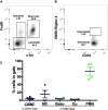
(A) Representative flow cytometric plot of the OSM+ gate. (B) Representative flow cytometric plots of the c-kit-FcεRI gate. (C) Quantification of the relative representation of different cell types among the OSM+ cells. Mast cells (MC) were 15.7±9.6%, basophils (Baso) were 3.7±1.5%, eosinophils (Eo) were 2.3±.9% and neutrophils (PMN) were 73.3±10.6% within the OSM+ gate, n=4. OSM+ cells were 4.6±1.9% of the CD45+ cells.
Figure 4
Figure 4. GM-CSF was elevated in nasal polyps and induced neutrophil derived OSM
(A) Blood neutrophils were stimulated with the indicated concentrations of GM-CSF, FSTL1 or both and levels of OSM protein in the culture supernatant were measured; (n=7–13, * p< .05, * p< .001, Kruskal-Wallis test). (B) Fully differentiated NHBE cells were either unstimulated, or stimulated with HKSA, and levels of GM-CSF protein in the cell culture supernatants were measured. HKSA treated NHBE released more GM-CSF, 21.3± 6.9pg/mL, than control NHBE, .87±.87 (n=4, p< .05, Mann-Whitney U test). (C) GM-CSF protein, 2.6±.93pg/mg total protein, was elevated in nasal polyps compared to control UT, .68±.42pg/mg total protein, (n=16–24, p< 05, Mann-Whitney U test). (D) FSTL1 protein, .60±.47ng/mL, was elevated in nasal polyps compared to control UT, .80±.33, (n=15–23, p=.31, Mann-Whitney U test). (E) OSM (alexa 488), GM-CSF (alexa 568) and neutrophil elastase (alexa 647) colocalized in nasal polyps (n=4).
Figure 5
Figure 5. OSM producing neutrophils did not express a classical N1 phenotype
(A) OSM+ neutrophils contained a distinct population of Arg1+MPOhi cells, representative data. (B) 3.7±1.3% of cells in the CD45 gate were OSM+; within the OSM+ gate, 56.0±8.2% of the cells were CD16+IL-5R− neutrophils. Within the neutrophil gate 72.56± 11.9% of the cells were Arg1+MPOhi, and 22.1± 8.0% were Arg1MPOlo, n=5. (C) Blood neutrophils were either left untreated, or treated under N1 polarizing conditions (LPS/IFNγ), N2 polarizing conditions (IL-4/IL-13) or with GM-CSF. GM-CSF treated neutrophils secreted elevated levels of OSM into the cell culture supernatants, while N1 and N2 polarizing conditions did not induce OSM, (n=4–7, p< .01, Kruskal-Wallis test). (D) Levels of MRC1 mRNA were elevated in neutrophils stimulated with GM-CSF either alone or together with FSTL1, (n=3–7, p< .05, Kruskal-Wallis test).
Figure 6
Figure 6. OSM was elevated in asthmatic patients
(A) Bronchial biopsies from control patients, moderate asthmatics and severe asthmatics were stained for OSM in green and neutrophil elastase in red, n=4–6. (B) Counts were obtained of neutrophils and OSM+ cells; none of the control biopsies had any OSM+ cells, and of the OSM+ cells 35± 21.8% were neutrophils in moderate asthmatics, and 52.1± 15.9% were neutrophils in severe asthmatics. (C) Levels of OSM in the sputum of asthmatic patients, 26.9± 9.5pg were elevated compared to control patients 3.9± 2.6pg, (n=11–12, p< .05, Mann-Whitney U test).
Figure 7
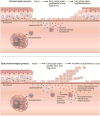
Figure 7. Proposed mechanism of OSM mediated barrier dysfunction in mucosal disease
Under normal circumstances, when epithelium is injured, neutrophils are recruited to the site of injury and potentially, transiently make OSM to promote the early stages of repair. Due to transient OSM expression, once the epithelial cells become contact inhibited they are able to enter the later stages of repair and redifferentiate back into functional epithelium. However, under pathogenic conditions we hypothesize that neutrophils are recruited to the injury site, and once at the site of injury the neutrophils are converted into an alternative phenotype, potentially N2, that makes both OSM and GM-CSF. The GM-CSF alone is sufficient to induce production of OSM in neutrophils, and will also contribute to long-term survival of OSM producing neutrophils that may prevent late stage repair, causing a long-term state of barrier dysfunction.

References

    1. Holgate ST, Lackie P, Wilson S, Roche W, Davies D. Bronchial epithelium as a key regulator of airway allergen sensitization and remodeling in asthma. American journal of respiratory and critical care medicine. 2000;162:S113–117. - PubMed
    1. Xiao C, et al. Defective epithelial barrier function in asthma. The Journal of allergy and clinical immunology. 2011;128:549–556. e541–512. - PubMed
    1. Dejima K, Randell SH, Stutts MJ, Senior BA, Boucher RC. Potential role of abnormal ion transport in the pathogenesis of chronic sinusitis. Archives of otolaryngology–head & neck surgery. 2006;132:1352–1362. - PubMed
    1. Bernstein JM, Gorfien J, Noble B, Yankaskas JR. Nasal polyposis: immunohistochemistry and bioelectrical findings (a hypothesis for the development of nasal polyps) The Journal of allergy and clinical immunology. 1997;99:165–175. - PubMed
    1. Kern RC, et al. Perspectives on the etiology of chronic rhinosinusitis: an immune barrier hypothesis. American journal of rhinology. 2008;22:549–559. - PMC - PubMed

MeSH terms

Substances