

Adaptation to Leftward Shifting Prisms Alters Motor Interhemispheric Inhibition
- PMID: 27993820
- PMCID: PMC6248503
- DOI: 10.1093/cercor/bhw386
Adaptation to Leftward Shifting Prisms Alters Motor Interhemispheric Inhibition
Abstract
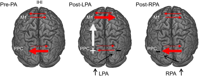
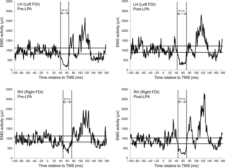
Adaptation to rightward shifting prisms (rightward prism adaptation, RPA) ameliorates neglect symptoms in patients while adaptation to leftward shifting prisms (leftward prism adaptation, LPA) induces neglect-like behaviors in healthy subjects. It has been hypothesized that prism adaptation (PA) modulates interhemispheric balance between the parietal cortices by inhibiting the posterior parietal cortex (PPC) contralateral to the prismatic deviation, but PA's effects on interhemispheric inhibition (IHI) have not been directly investigated. Since there are hyper-excitable connections between the PPC and primary motor cortex (M1) in the left hemisphere of neglect patients, we reasoned that LPA might mimic right hemisphere lesions by reducing parietal IHI, hyper-exciting the left PPC and PPC-M1 connections, and in turn altering IHI at the motor level. Namely, we hypothesized that LPA would increase IHI from the left to the right M1. We examined changes in left-to-right and right-to-left IHI between the 2 M1s using the ipsilateral silent period (iSP) (Meyer et al. 1995) before and after either LPA or RPA. The iSP was significantly longer after LPA but only from left-to-right and it did not change at all after RPA. This is the first physiological demonstration that LPA alters IHI in the healthy brain.
Keywords: TMS; ipsilateral silent period; prism adaptation; visuospatial neglect.
© The Author 2016. Published by Oxford University Press. All rights reserved. For Permissions, please e-mail: journals.permissions@oup.com.
Figures
Similar articles
-
Choosing Sides: Impact of Prismatic Adaptation on the Lateralization of the Attentional System.Front Psychol. 2022 Jun 23;13:909686. doi: 10.3389/fpsyg.2022.909686. eCollection 2022. Front Psychol. 2022. PMID: 35814089 Free PMC article. Review.
-
The asymmetrical effect of leftward and rightward prisms on intact visuospatial cognition.Cortex. 2017 Dec;97:23-31. doi: 10.1016/j.cortex.2017.09.015. Epub 2017 Oct 3. Cortex. 2017. PMID: 29078083 Free PMC article. Clinical Trial.
-
Paired-Pulse Parietal-Motor Stimulation Differentially Modulates Corticospinal Excitability across Hemispheres When Combined with Prism Adaptation.Neural Plast. 2016;2016:5716179. doi: 10.1155/2016/5716179. Epub 2016 Jun 22. Neural Plast. 2016. PMID: 27418979 Free PMC article.
-
Variation in left posterior parietal-motor cortex interhemispheric facilitation following right parietal continuous theta-burst stimulation in healthy adults.Neuroscience. 2016 Aug 25;330:229-35. doi: 10.1016/j.neuroscience.2016.05.056. Epub 2016 Jun 4. Neuroscience. 2016. PMID: 27267243
-
Modulation of visual attention by prismatic adaptation.Neuropsychologia. 2016 Nov;92:31-41. doi: 10.1016/j.neuropsychologia.2016.06.022. Epub 2016 Jun 21. Neuropsychologia. 2016. PMID: 27342255 Review.
Cited by
-
Choosing Sides: Impact of Prismatic Adaptation on the Lateralization of the Attentional System.Front Psychol. 2022 Jun 23;13:909686. doi: 10.3389/fpsyg.2022.909686. eCollection 2022. Front Psychol. 2022. PMID: 35814089 Free PMC article. Review.
-
Prismatic Adaptation Modulates Oscillatory EEG Correlates of Motor Preparation but Not Visual Attention in Healthy Participants.J Neurosci. 2018 Jan 31;38(5):1189-1201. doi: 10.1523/JNEUROSCI.1422-17.2017. Epub 2017 Dec 18. J Neurosci. 2018. PMID: 29255004 Free PMC article.
-
Improvement of phonemic fluency following leftward prism adaptation.Sci Rep. 2021 Mar 31;11(1):7313. doi: 10.1038/s41598-021-86625-0. Sci Rep. 2021. PMID: 33790347 Free PMC article.
-
Virtual reality-based sensorimotor adaptation shapes subsequent spontaneous and naturalistic stimulus-driven brain activity.Cereb Cortex. 2023 Apr 25;33(9):5163-5180. doi: 10.1093/cercor/bhac407. Cereb Cortex. 2023. PMID: 36288926 Free PMC article.
-
Neural Mechanisms of Prism Adaptation in Healthy Adults and Individuals with Spatial Neglect after Unilateral Stroke: A Review of fMRI Studies.Brain Sci. 2021 Nov 5;11(11):1468. doi: 10.3390/brainsci11111468. Brain Sci. 2021. PMID: 34827467 Free PMC article. Review.
References
-
- Bagattini C, Mele S, Brignani D, Savazzi S. 2015. No causal effect of left hemisphere hyperactivity in the genesis of neglect-like behavior. Neuropsychologia. 72:12–21. - PubMed
-
- Bartolomeo P. 2015. Spatially biased decisions: toward a dynamic interactive model of visual neglect In: Tracy JI, Hampstead BM, Sathian K, editors.. Plasticity of cognition in neurologic disorders. Oxford: Oxford University Press; p. 299–322.
-
- Bartolomeo P, Chokron S. 1999. Left unilateral neglect or right hyperattention? Neurology. 53:2023–2027. - PubMed
-
- Bartolomeo P, D'Erme P, Perri R, Gainotti G. 1998. Perception and action in hemispatial neglect. Neuropsychologia. 36:227–237. - PubMed
Publication types
MeSH terms
LinkOut - more resources
Full Text Sources
Other Literature Sources
Miscellaneous
