. 2017 Mar 1;37(6):e00401-16.
doi: 10.1128/MCB.00401-16. Print 2017 Mar 15.

Human Apurinic/Apyrimidinic Endonuclease (APE1) Is Acetylated at DNA Damage Sites in Chromatin, and Acetylation Modulates Its DNA Repair Activity

Affiliations

Human Apurinic/Apyrimidinic Endonuclease (APE1) Is Acetylated at DNA Damage Sites in Chromatin, and Acetylation Modulates Its DNA Repair Activity

Shrabasti Roychoudhury et al. Mol Cell Biol. .

Abstract

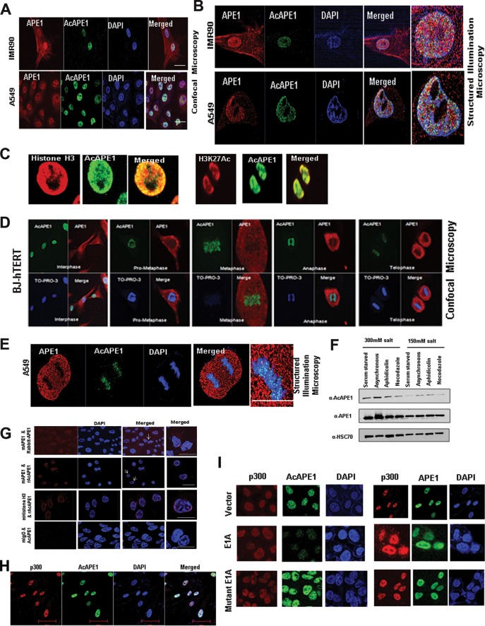
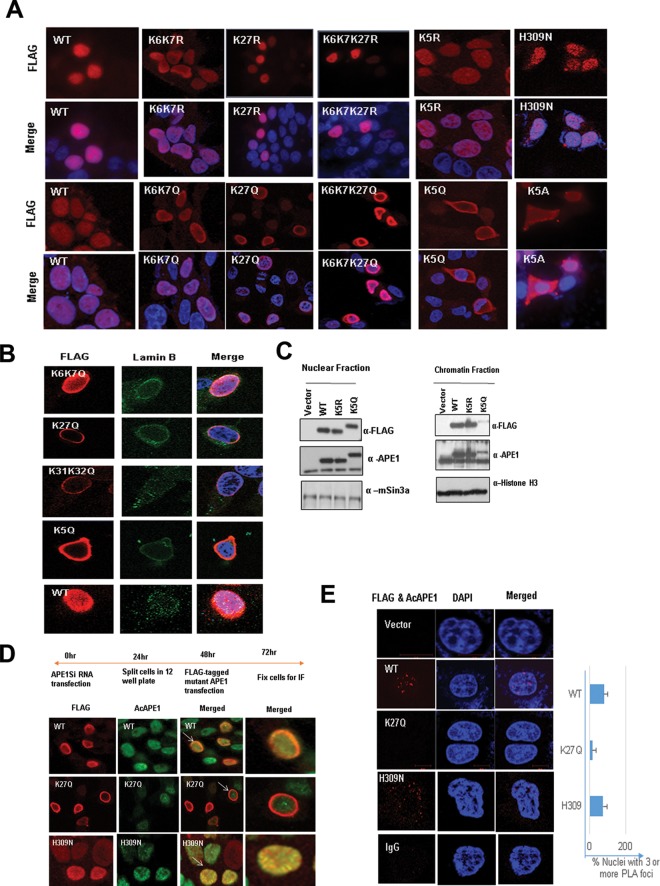
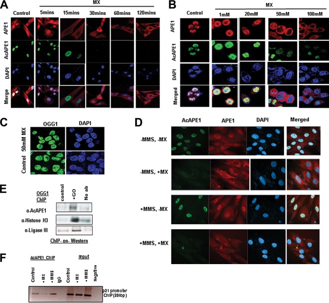
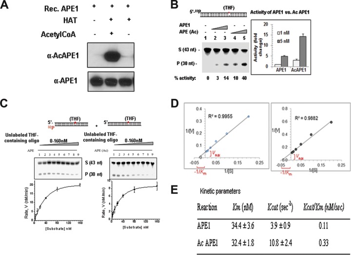
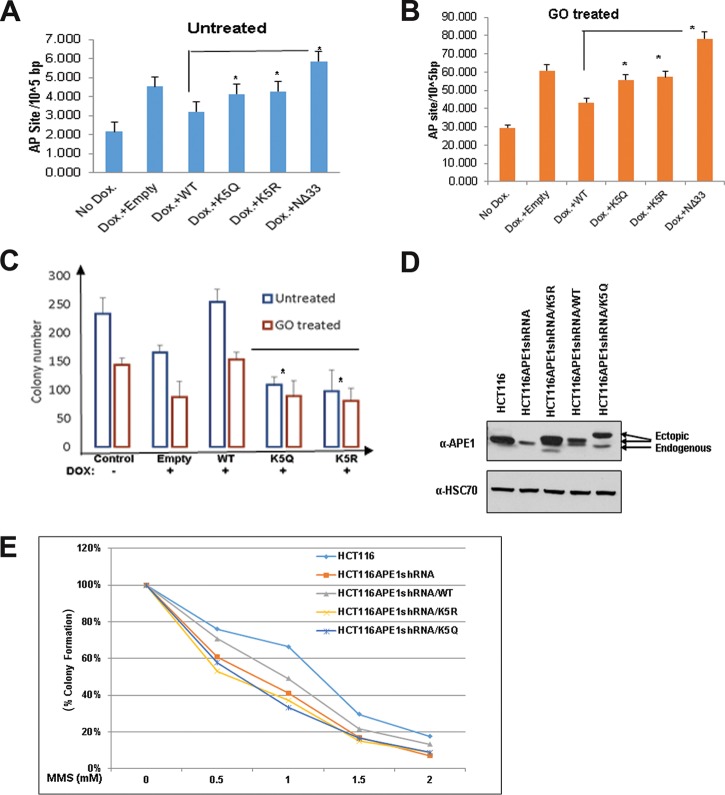
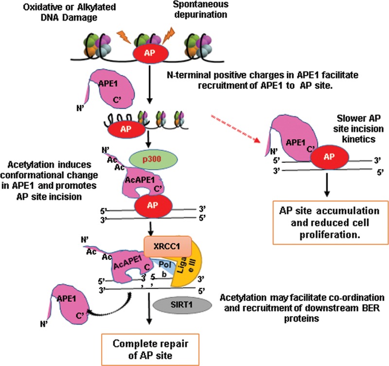
Apurinic/apyrimidinic (AP) sites, the most frequently formed DNA lesions in the genome, inhibit transcription and block replication. The primary enzyme that repairs AP sites in mammalian cells is the AP endonuclease (APE1), which functions through the base excision repair (BER) pathway. Although the mechanism by which APE1 repairs AP sites in vitro has been extensively investigated, it is largely unknown how APE1 repairs AP sites in cells. Here, we show that APE1 is acetylated (AcAPE1) after binding to the AP sites in chromatin and that AcAPE1 is exclusively present on chromatin throughout the cell cycle. Positive charges of acetylable lysine residues in the N-terminal domain of APE1 are essential for chromatin association. Acetylation-mediated neutralization of the positive charges of the lysine residues in the N-terminal domain of APE1 induces a conformational change; this in turn enhances the AP endonuclease activity of APE1. In the absence of APE1 acetylation, cells accumulated AP sites in the genome and showed higher sensitivity to DNA-damaging agents. Thus, mammalian cells, unlike Saccharomyces cerevisiae or Escherichia coli cells, require acetylation of APE1 for the efficient repair of AP sites and base damage in the genome. Our study reveals that APE1 acetylation is an integral part of the BER pathway for maintaining genomic integrity.

Keywords: AP site; APE1; DNA damage; acetylation; base excision repair; endogenous DNA damage.


Figures

FIG 1
FIG 1
AcAPE1 is exclusively associated with chromatin and remains bound to the condensed chromosomes. (A and B) Asynchronous normal lung fibroblast IMR90 cells and lung adenocarcinoma A549 cells were immunostained with anti-APE1 and anti-AcAPE1 Abs, counterstained with DAPI, and visualized by confocal microscopy and 3D SIM. (C) Colocalization of AcAPE1 with histone H3 or active enhancer-specific histone marker acetylated H3K27 (H3K27Ac). (D) BJ-hTERT cells were serum starved for 72 h and then fixed at different time points. Cells were immunostained with anti-APE1 and anti-AcAPE1 Abs and counterstained with anti-TO-PRO-3 iodide Ab. (E) Mitotic A549 cells were immunostained with anti-APE1 and anti-AcAPE1 and visualized by 3D SIM. (F) BJ-hTERT cells were either serum starved for 72 h (G0/G1 phase), treated with nocodazole (mitotic cells) or aphidicolin (G1/S phase synchronized cells), or untreated, and whole-cell extracts were isolated using 150 mM or 300 mM salt-containing lysis buffer. Western blot analysis for anti-APE1 and anti-AcAPE1 levels was performed. Anti-HSC70 was used as loading control. (G) A proximal ligation assay was performed with mouse anti-APE1 and rabbit anti-APE1 (mAPE1 & Rabbit-APE1), mouse anti-mouse APE1 and rabbit anti-AcAPE1 (mAPE1 & rAcAPE1), and rabbit anti-AcAPE1 and mouse anti-histone H3 (mHistone H3 & rAcAPE1) to confirm the chromatin association of AcAPE1. Mouse IgG (mIgG) and rabbit anti-AcAPE1 were used as a control. At least 50 cells were counted for PLA foci. (H) Colocalization of p300 and AcAPE1 on chromatin (DAPI). (I) HCT116 cells were transfected with E1A and mutant E1A, and at 48 h after transfection, IF was performed. Cells were immunostained with anti-p300 and anti-APE1 or anti-AcAPE1 and counterstained with DAPI.
FIG 2
FIG 2
Positive charges of acetylable Lys residues but not their acetylation are essential for chromatin binding of APE1. (A) Cells expressing different mutants with Lys site-specific APE1 mutations were immunostained with anti-FLAG Ab (rows labeled FLAG) and counterstained with DAPI (rows labeled Merge). (B) The subcellular localization of acetylation site mutants was analyzed by immunostaining; cells expressing mutants with different Lys site-specific APE1 mutations were stained with anti-FLAG Ab or anti-lamin B Ab and counterstained with DAPI. (C) Western blot analysis of soluble nuclear and chromatin extracts for FLAG-tagged WT and mutant APE1 levels in cells ectopically expressing these proteins with anti-FLAG. Anti-APE1, anti-histone H3, and anti-mouse Sin3a (mSin3a) were used as controls. (D and E) Schematic overview of the experiment. APE1 was downregulated in HEK293T cells using APE1-specific siRNA (APE1Si RNA), and after 48 h, the cells were transfected with expression plasmids containing WT APE1 or K27Q or H309N mutant APE1. IF was performed using anti-FLAG and anti-AcAPE1 antibodies to check colocalization. At least 50 FLAG-positive cells were counted for colocalization. Cells indicated with arrows were further magnified in the “Merged” column. (E) (Left) PLA was performed using anti-FLAG and anti-AcAPE1 to examine the acetylation of WT APE1 and K27Q and H309N mutant APE1 in cells. (Right) At least 50 cells were counted, and the percentage of the PLA signal was plotted for each APE1 mutant.
FIG 3
FIG 3
APE1 is acetylated after binding to AP sites in the chromatin. (A) BJ-hTERT cells were treated with MX (50 mM) for the indicated times. IF was performed using anti-APE1 and anti-AcAPE1, and counterstaining with DAPI was used. (B) HCT116 cells were treated with various doses of MX for 30 min, IF was performed using anti-APE1 and anti-AcAPE1, and counterstaining with DAPI was used. (C) HCT116 cells were treated with 50 mM MX for 30 min, IF was performed using anti-OGG1, and counterstaining with DAPI was used. (D) BJ-hTERT cells pretreated with 50 mM MX for 30 min or not pretreated were exposed to MMS (2 mM) for 1 h. IF was performed using anti-APE1 and anti-AcAPE1, and counterstaining with DAPI was used. Confocal microscopy was used to visualize the AcAPE1 levels in control cells and cells treated with MMS or MX, or both. (E) ChIP with anti-OGG1 antibody followed by Western blotting (ChIP-on-Western) was performed to examine the association of AcAPE1 and ligase III on chromatin after induction of DNA damage with GO. (F) The association of AcAPE1 with the endogenous p21 promoter in control or MMS- or MX-treated cells was examined by promoter-directed ChIP using anti-AcAPE1.
FIG 4
FIG 4
Acetylation of APE1 enhances its AP endonuclease activity. (A) Recombinant (Rec.) APE1 was incubated with the p300 HAT domain either in the presence or the absence of acetyl-CoA, and Western blot analysis was performed with anti-APE1 and anti-AcAPE1 Abs to confirm the acetylation of APE1. (B) Incision of the THF (reduced AP site)-containing 43-mer duplex oligonucleotide (S, substrate) by APE1 and in vitro-acetylated APE1. nt, nucleotides; P, the cleaved product. (C and D) The values of the kinetic parameters Km and kcat were calculated by incubating 33 pM enzyme at 37°C for 3 min with substrates at various concentrations (0 to 160 nM). The enzyme kinetics data were fitted into a nonlinear least-squares regression to obtain Vmax and Km values by use of the Michaelis-Menten equation and SigmaPlot software. (E) Comparison of kinetic parameters between APE1 and AcAPE1.
FIG 5
FIG 5
APE1 acetylation enhances its stability on chromatin and its interaction with downstream BER proteins. (A) Colocalization of ligase III and AcAPE1 in A549 cells. Cells were immunostained with anti-ligase III and anti-AcAPE1 Abs. (B) WT or K5R mutant APE1-overexpressing HEK293T cells were treated with TSA-nicotinamide (NAM) for 6 h or not treated, and the nuclear extract was immunoprecipitated using anti-FLAG Ab and immunoblotted with anti-XRCC1 and anti-FLAG Abs. (C) A549 cells were fixed with paraformaldehyde before (top) or after treatment with Triton X-100 (0.5%) (middle) or Triton X-100 plus salt (100 mM KCl) (bottom) and immunostained with anti-APE1 or anti-AcAPE1 Abs and counterstained with DAPI. (D) Acetylation of APE1 induces a conformational change in APE1. The distinct intrinsic fluorescence emission spectra of APE1 and AcAPE1 at 280 nm are shown. A.U., absorbance units.
FIG 6
FIG 6
APE1 acetylation is essential for cell survival and/or cell proliferation, and the absence of APE1 acetylation sensitizes cells to DNA-damaging agents. (A and B) Endogenous APE1 was downregulated in HEK293TAPE1siRNA cells using Dox treatment. (A) FLAG-tagged WT APE1, acetylation-defective mutants (mutants with the mutation of Lys6, Lys7, Lys27, Lys31, and Lys32 to nonacetylable arginine [K5R] or glutamine [K5Q]), or NΔ33 mutants were further overexpressed in these cells. Cells were treated with glucose oxidase (100 ng/ml for 30 min) or not treated, and the number of AP sites was measured using an ARP kit. (B) Bar diagram representing the number of AP sites/105 bp in the presence of different APE1 mutants compared to that for the vector control. Error bars indicate means ± SDs (n = 3). (C) HEK293TAPE1siRNA cells were treated with DOX or not treated, and WT APE1 and K5R and K5Q mutant APE1 were ectopically expressed. Cells were treated with glucose oxidase (100 ng/ml for 30 min) or not treated, and a colony formation assay was performed. The bar diagram shows the number of colonies formed in the presence of the different APE1 mutants compared to that for the vector control. Error bars indicate means ± SDs (n = 3). (D and E) HCT116 cells constitutively expressing APE1 shRNA (HCT116APE1shRNA cells) were transfected with FLAG-tagged WT APE1 or acetylation-defective APE1 mutants (mutants with mutation of Lys6, Lys7, Lys27, Lys31, and Lys32 to nonacetylable arginine [K5R] or glutamine [K5Q]). (D) Western blot analysis was performed to examine APE1 levels using an anti-APE1 Ab. Anti-HSC70 was used as a loading control. (E) Sensitivity to damage was measured in HCT116APE1shRNA cells ectopically expressing different APE1 mutants. Cells were treated with increasing doses of MMS for 1 h, and a colony formation assay was performed.
FIG 7
FIG 7
Schematic model for regulation of AP site repair in cells by acetylation of APE1 via the BER pathway.

Similar articles

Cited by

References

    1. Lindahl T, Andersson A. 1972. Rate of chain breakage at apurinic sites in double-stranded deoxyribonucleic acid. Biochemistry 11:3618–3623. doi:10.1021/bi00769a019. - DOI - PubMed
    1. Lindahl T. 1993. Instability and decay of the primary structure of DNA. Nature 362:709–715. doi:10.1038/362709a0. - DOI - PubMed
    1. Loeb LA, Preston BD. 1986. Mutagenesis by apurinic/apyrimidinic sites. Annu Rev Genet 20:201–230. doi:10.1146/annurev.ge.20.120186.001221. - DOI - PubMed
    1. Tornaletti S, Maeda LS, Hanawalt PC. 2006. Transcription arrest at an abasic site in the transcribed strand of template DNA. Chem Res Toxicol 19:1215–1220. doi:10.1021/tx060103g. - DOI - PubMed
    1. Tell G, Quadrifoglio F, Tiribelli C, Kelley MR. 2009. The many functions of APE1/Ref-1: not only a DNA repair enzyme. Antioxid Redox Signal 11:601–620. doi:10.1089/ars.2008.2194. - DOI - PMC - PubMed

Publication types

LinkOut - more resources