. 2017 Feb 3;292(5):1559-1572.
doi: 10.1074/jbc.M116.759811. Epub 2016 Dec 19.

Stabilization of Nucleotide Binding Domain Dimers Rescues ABCC6 Mutants Associated with Pseudoxanthoma Elasticum

Affiliations

Stabilization of Nucleotide Binding Domain Dimers Rescues ABCC6 Mutants Associated with Pseudoxanthoma Elasticum

Yanchao Ran et al. J Biol Chem. .

Abstract

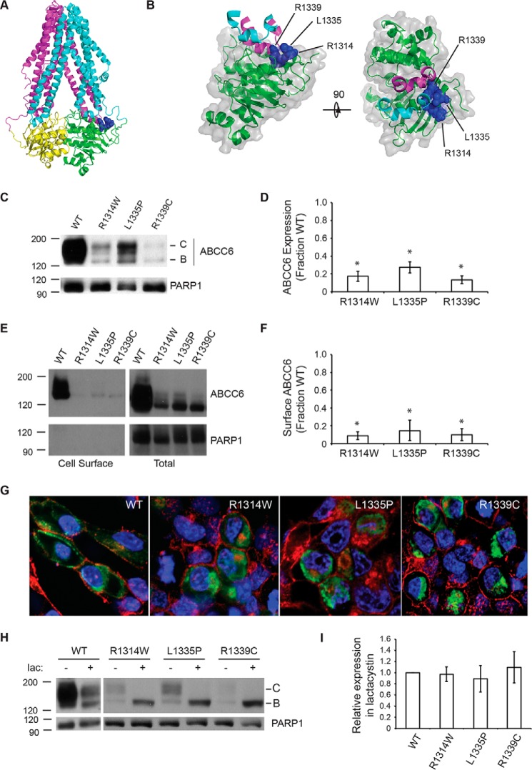
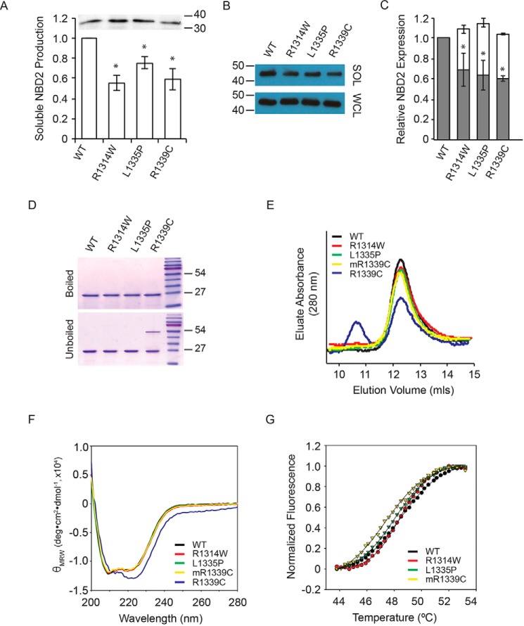
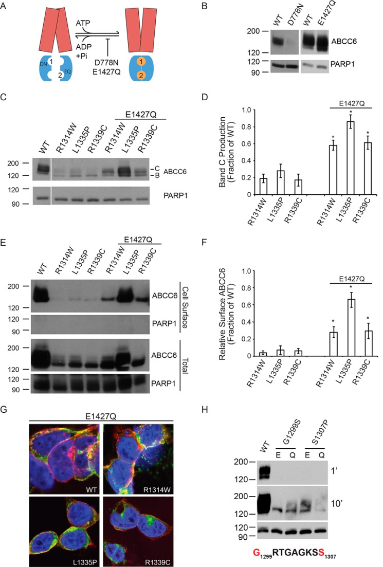
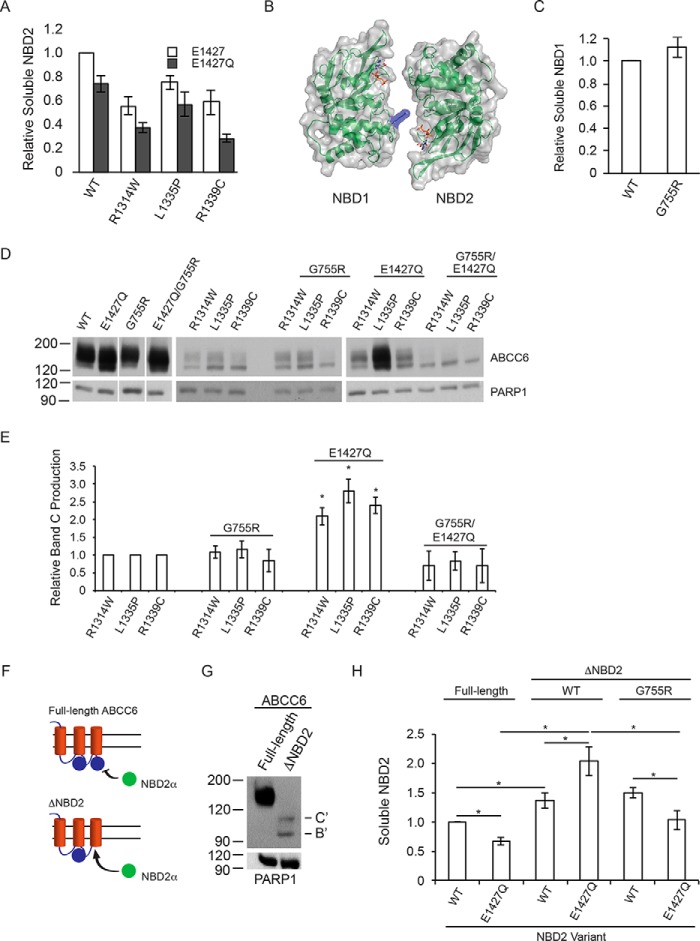
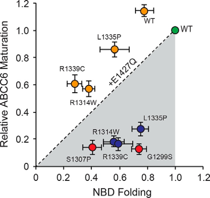
ABC transporters are polytopic membrane proteins that utilize ATP binding and hydrolysis to facilitate transport across biological membranes. Forty-eight human ABC transporters have been identified in the genome, and the majority of these are linked to heritable disease. Mutations in the ABCC6 (ATP binding cassette transporter C6) ABC transporter are associated with pseudoxanthoma elasticum, a disease of altered elastic properties in multiple tissues. Although ∼200 mutations have been identified in pseudoxanthoma elasticum patients, the underlying structural defects associated with the majority of these are poorly understood. To evaluate the structural consequences of these missense mutations, a combination of biophysical and cell biological approaches were applied to evaluate the local and global folding and assembly of the ABCC6 protein. Structural and bioinformatic analyses suggested that a cluster of mutations, representing roughly 20% of the patient population with identified missense mutations, are located in the interface between the transmembrane domain and the C-terminal nucleotide binding domain. Biochemical and cell biological analyses demonstrate these mutations influence multiple steps in the biosynthetic pathway, minimally altering local domain structure but adversely impacting ABCC6 assembly and trafficking. The differential impacts on local and global protein structure are consistent with hierarchical folding and assembly of ABCC6. Stabilization of specific domain-domain interactions via targeted amino acid substitution in the catalytic site of the C-terminal nucleotide binding domain restored proper protein trafficking and cell surface localization of multiple biosynthetic mutants. This rescue provides a specific mechanism by which chemical chaperones could be developed for the correction of ABCC6 biosynthetic defects.

Keywords: ABC transporter; abcc6; biosynthesis; calcification; protein stability; protein structure; pseudoxanthoma elasticum.


Conflict of interest statement

The authors declare that they have no conflicts of interest with the contents of this article

Figures

FIGURE 1.
FIGURE 1.
ABCC6 biosynthesis and trafficking. The NBD2 mutations, R1314W, L1335P, and R1339C, were assessed for their impacts on ABCC6 structure and biosynthesis. A, a schematic representation of an ABCC6 homology model is shown. The core TMDs are colored cyan and magenta, and the NBDs are colored green and yellow. The Arg-1314, Leu-1335, and Arg-1339 side chains are shown as blue spheres. B, a schematic and surface representation of NBD2 and the contacting intracellular loops is shown. Intracellular loop four, from TMD1, is shown in cyan, and ICL5 (intracellular loop 5) from TMD2 is shown in magenta. The surface of NBD2 associated with the Arg-1314, Leu-1335, and Arg-1339 residues is shown in blue. C, a representative Western blot of HEK293 cells expressing the wild type, R1314W, L1335P, and R1339C mutants is shown. D, densitometric analysis of Western blots of steady state ABCC6 is shown. E, representative Western blotting after cell surface biotinylation is shown. F, densitometric analysis of cell surface resident ABCC6 is shown. G, representative confocal immunofluorescence images of HEK293 cells expressing the wild type and mutant ABCC6 proteins are shown. ABCC6 is stained in green, WGA is shown in red, and DAPI is shown in blue. H, a representative Western blot of HEK293 cells expressing ABCC6 in the presence and absence of lactacystin is shown. I, densitometric analysis of Western blots of ABCC6 after lactacystin treatment is shown normalized to the expression of wildtype ABCC6. Western blots and immunofluorescence images are representative of n ≥ 4 independent experiments. The identities of band B and C are indicated in C and H. PARP1 is shown as a loading control in C, E, and F. Data shown are summary or representative of n ≥ 3 independent experiments. Quantified data are mean ± S.D. *, p < 0.01 using ANOVA with Tukey's post hoc test.
FIGURE 2.
FIGURE 2.
NBD2 folding and structure. The effects of the three NBD2 mutants were evaluated on the isolated NBD2 protein. A, NBD2 folding was assessed using the β-galactosidase structural complementation assay and normalized to the wild type NBD2 signal. Inset, a representative Western blot of total NBD2 protein expression is shown. B, Western blots of the whole cell lysate (WCL) and soluble fractions of NBD2 protein expressed in E. coli are shown. C, quantification of soluble NBD2 protein expression is shown. The open bars indicate the whole cell lysate, and the gray bars indicate the soluble fraction remaining after centrifugation. D, representative Coomassie Blue-stained gels are shown before and after boiling in the presence of nucleotide and reductant. E, chromatographs of the purified NBD2 proteins are shown after separation by analytical gel filtration. F, circular dichroism spectra of the purified NBD2 proteins are shown. G, SYPRO Orange thermal denaturation transitions are shown for the purified NBD2 proteins. Data shown are the summary or representative of n ≥ 3 independent experiments. Quantified data are the mean ± S.D. *, p < 0.01 using ANOVA with Tukey's post hoc test.
FIGURE 3.
FIGURE 3.
Rescued ABCC6 folding by domain-domain stabilization. The catalytic E1427Q mutation was used to stabilize the ATP-bound NBD dimer and to assess the rescue of ABCC6 folding. A, a schematic showing the putative domain-domain stabilization by the E1427Q mutant is shown. The catalytic mutation, E1427Q, putatively blocks ATP hydrolysis at the NBD2 Walker A/B composite site. The TMDs are shown in red, the NBDs are shown in blue, and the ATP is shown as orange spheres. The ATP molecules are numbered based on their association with the NBD1 or NBD2 Walker A/B sequences. B, representative Western blots of the NBD1 and NBD2 catalytic mutants in the otherwise wild type ABCC6 background are shown. C, representative Western blots of the E1427Q double mutants are shown. D, densitometric analysis of Western blots of steady state ABCC6 is shown. E, representative Western blots of cell surface biotinylated ABCC6 are shown. F, densitometric analysis of Western blots of steady state ABCC6 is shown. G, confocal immunofluorescence images of the E1427Q mutants are shown. ABCC6 is shown in green, WGA is shown in red, and DAPI is shown in blue. Yellow indicates colocalization of the ABCC6 and agglutinin staining. H, representative Western blots of Walker A ATP-binding site mutations are shown with Glu-1427 (E) or E1427Q (Q). Western blots and immunofluorescence images are representative of n ≥ 4 independent experiments. The identities of band B and C are indicated in C. PARP1 is shown as a loading control in B, C, E, and H. Data shown are summary or representative of n ≥ 3 independent experiments. Quantified data are mean ± S.D. *, p < 0.01 using ANOVA with Tukey's post hoc test.
FIGURE 4.
FIGURE 4.
NBD structure, dimerization, and biosynthetic correction of ABCC6. The role of NBD dimerization in the E1427Q-mediated ABCC6 rescue was assessed. A, soluble NBD2, assessed using the β-galactosidase assay is shown normalized to the wild type NBD2 signal. B, a schematic and surface representation of the ABCC6 NBD heterodimer is shown with the G755R substitution shown in blue. C, soluble NBD1 protein, measured using the β-galactosidase assay is shown normalized to wild type NBD1 signal. D, representative Western blots of the wild type, E1427Q, and G755R mutant ABCC6 proteins are shown with and without the NBD2 mutations. E, densitometric analysis of Western blots is shown for the NBD2, G755R and E1427Q mutants. F, a schematic showing the trans complementation of NBD2 co-expressed with full-length or truncated forms of ABCC6 is shown. G, a representative blot of the biosynthesis of the ΔNBD2 protein is shown. The core and glycosylated forms are indicated with B and C, respectively, with B′ and C′ indicated the core and fully glycosylated species of the truncated ABCC6 protein. H, co-expression of NBD2 with the ΔNBD2 protein is assessed by the β-galactosidase assay. Data shown are summary or representative of n ≥ 3 independent experiments. Quantified data are mean ± S.D. *, p < 0.01 using ANOVA with Tukey's post hoc test.
FIGURE 5.
FIGURE 5.
Relationship between NBD and full-length ABCC6 folding. The impact of the NBD2 mutations on the production of full-length and soluble NBD2 proteins is shown. NBD2 folding, measured as a function of soluble NBD2 production is plotted along the x axis. Full-length ABCC6 protein production, measured as the relative quantity of band C, fully glycosylated protein, is plotted on the y axis. Values for both are normalized against the wild type signal, green. The R1314W, L1335P, and R1339C single mutants are shown in red; the E1427Q variant proteins are shown in orange. The dashed line represents the unity relationship between NBD folding and ABCC6 maturation.

References

    1. Le Saux O., Urban Z., Tschuch C., Csiszar K., Bacchelli B., Quaglino D., Pasquali-Ronchetti I., Pope F. M., Richards A., Terry S., Bercovitch L., de Paepe A., and Boyd C. D. (2000) Mutations in a gene encoding an ABC transporter cause pseudoxanthoma elasticum. Nat. Genet. 25, 223–227 - PubMed
    1. Jiang Q., Endo M., Dibra F., Wang K., and Uitto J. (2009) pseudoxanthoma elasticum is a metabolic disease. J. Invest. Dermatol. 129, 348–354 - PMC - PubMed
    1. Goodman R. M., Smith E. W., Paton D., Bergman R. A., Siegel C. L., Ottesen O. E., Shelley W. M., Pusch A. L., and McKusick V. A. (1963) Pseudoxanthoma elasticum: a clinical and histopathological study. Medicine 42, 297–334 - PubMed
    1. Marconi B., Bobyr I., Campanati A., Molinelli E., Consales V., Brisigotti V., Scarpelli M., Racchini S., and Offidani A. (2015) Pseudoxanthoma elasticum and skin: clinical manifestations, histopathology, pathomechanism, perspectives of treatment. Intractable Rare Dis. Res. 4, 113–122 - PMC - PubMed
    1. Jansen R. S., Duijst S., Mahakena S., Sommer D., Szeri F., Váradi A., Plomp A., Bergen A. A., Oude Elferink R. P., Borst P., and van de Wetering K. (2014) ABCC6-mediated ATP secretion by the liver is the main source of the mineralization inhibitor inorganic pyrophosphate in the systemic circulation-brief report. Arterioscler. Thromb. Vasc. Biol. 34, 1985–1989 - PMC - PubMed

Publication types

MeSH terms

Substances

LinkOut - more resources