. 2017 Jan 27;292(4):1438-1448.
doi: 10.1074/jbc.M116.759886. Epub 2016 Dec 19.

Trp-Asp (WD) Repeat Domain 1 Is Essential for Mouse Peri-implantation Development and Regulates Cofilin Phosphorylation

Affiliations

Trp-Asp (WD) Repeat Domain 1 Is Essential for Mouse Peri-implantation Development and Regulates Cofilin Phosphorylation

Yi Xiao et al. J Biol Chem. .

Abstract

Trp-Asp (WD) repeat domain 1 (WDR1) is a highly conserved actin-binding protein across all eukaryotes and is involved in numerous actin-based processes by accelerating Cofilin severing actin filament. However, the function and the mechanism of WDR1 in mammalian early development are still largely unclear. We now report that WDR1 is essential for mouse peri-implantation development and regulates Cofilin phosphorylation in mouse cells. The disruption of maternal WDR1 does not obviously affect ovulation and female fertility. However, depletion of zygotic WDR1 results in embryonic lethality at the peri-implantation stage. In WDR1 knock-out cells, we found that WDR1 regulates Cofilin phosphorylation. Interestingly, WDR1 is overdosed to regulate Cofilin phosphorylation in mouse cells. Furthermore, we showed that WDR1 interacts with Lim domain kinase 1 (LIMK1), a well known phosphorylation kinase of Cofilin. Altogether, our results provide new insights into the role and mechanism of WDR1 in physiological conditions.

Keywords: Limk1; WDR1; actin; cofilin; development; embryonic development; mouse genetics; phosphorylation.


Figures

FIGURE 1.
FIGURE 1.
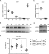
Maternal knock-out of Wdr1. A, Wdr1 transcripts were detected in 50 GV oocytes, MII oocytes, zygotes (1-C), two-cell (2-C), morula (MO), and blastocyst (BL) stage embryos by quantitative RT-PCR. RNA expression in GV oocytes was set as 1. Data are represented as mean ± S.D. from three independent experiments. B, expression of WDR1 protein in oocytes and preimplantation embryos (n = 50) were analyzed by Western blotting analysis with α-tubulin as a loading control. C, Wdr1 transcription was detected in 50 control or knock-out GV oocytes by real-time RT-PCR. RNA expression in control GV oocytes was set as 1. Error bars represent S.D. D, WDR1 protein was detected in 20 control or knock-out GV oocytes using GAPDH as a loading control. E, litter sizes of Wdr1f/f, Wdr1f/+, and Wdr1f/f; Zp3-cre females after mating with Wdr1f/f males or Wdr1f/f; Zp3-cre females after mating with Wdr1f/− males are presented as mean ± S.D. All litter sizes for each genotype were obtained from at least 3 females. ***, p < 0.001; NS, not significant.
FIGURE 2.
FIGURE 2.
Wdr1 KO embryos in peri-implantation development. A, after mating with Wdr1f/− males, oocyte specific knock-out females were injected with Chicago blue. Then, the uteruses were picked up at E5.5 and imaged by differential interference contrast. Scale bar, 5 mm. B, deciduas with embryos from A were frozen and sectioned (8 μm). The sections were stained by H&E and imaged. Two types such as (a) and (b) were observed. The percentage of the two types was counted from three pregnant females. Statistical results are represented as mean ± S.D. Scale bar, 50 μm. C, oocyte-specific Wdr1 KO females were mated with Wdr1f/− males. At E3.5, blastocysts were flushed from the uterus and imaged by differential interference contrast. Scale bar, 40 μm. D, blastocysts obtained from C were cultured in vitro for 3 days and treated with EdU for 2 h before being fixed. The outgrowth was stained for EdU (red) according to the protocol provided by the manufacturer. Nuclear DNA was stained by Hoechst 33342 (blue). Scale bar, 200 μm. E, Wdr1f/− (control, CTL) and Wdr1−/− (knock-out, KO) embryos were genotyped with specific primers.
FIGURE 3.
FIGURE 3.
Characterization of Wdr1 KO ES cells. A, total RNA samples from Wdr1f/− (control, CTL) and Wdr1−/− (knock-out, KO) ES were subjected to real-time RT-PCR to examine Wdr1 transcription. Gapdh mRNA was used as a control. B, WDR1 and pluripotential markers NANOG, OCT4, and SOX2 were analyzed by Western blotting in CTL and KO ES cell lines, with GAPDH as a loading control. C, two KO and two CTL ES cell lines were cultured in a 12-well plate and the cells were counted every 24 h. Three wells for each cell line were examined at each time point. D, CTL and KO ES cells were labeled with EGFP and injected into normal blastocysts. After culturing 3 days, the embryos were examined by microscopy. Scale bar, 20 μm. E, Pax6, T, and Sox17 transcription were examined by real-time RT-PCR in CTL and KO ES (d0) and 3 days EB (d3). Gapdh mRNA was used as a control. F, both CTL and Wdr1 KO ES cells were fixed and incubated with a primary antibody to Cofilin. Then, these cells were incubated with FITC-tagged second antibody (Cofilin, green), Alexa Fluor® 546 Phalloidin (F-actin, red), and Hoechst 33342 (DNA, blue), and imaged. Scale bar, 20 μm. G, P-Cofilin and total Cofilin were detected by immunoblotting in CTL and Wdr1 KO ES cells. GAPDH was used as a loading control. H, G-actin and F-actin were separated in control and Wdr1 KO ES cell lines and probed with mouse anti-actin antibody.
FIGURE 4.
FIGURE 4.
Characterization of Wdr1 mutant MEF cells. A, WDR1 was analyzed by Western blotting in ES and MEF cells, with GAPDH as a loading control. B, Wdr1 mRNA was detected by real-time RT-PCR in Wdr1+/−; Cre-ERTM (control, CTL) and Wdr1f/−; Cre-ERTM (iKO) MEF cells after being treated with tamoxifen for 72 h. C, immunoblot analyzed CTL and iKO MEF cells using WDR1 and GAPDH (loading control) antibodies. D, CTL and iKO MEF cells were stained by Alexa Fluor® 546 Phalloidin (F-actin, red) and Hoechst 33342 (DNA, blue), and imaged. Scale bar, 80 μm. E, G-actin and F-actin in CTL and iKO MEF cells were separated and probed by Western blotting with actin antibody. F, P-Cofilin, total-Cofilin, and WDR1 were detected in CTL and iKO MEF cells by Western blotting, using GAPDH as a loading control. G, after being treated with tamoxifen for 72 h, CTL and iKO MEF cells were digested, stained with PI, and examined by flow cytometry.
FIGURE 5.
FIGURE 5.
The regulation of WDR1 on Cofilin phosphorylation. A, Wdr1+/−; Cre-ERTM and Wdr1f/−; Cre-ERTM MEF cells were lysed after tamoxifen treatment at 0, 24, 48, and 72 h. Immunoblots of the samples were probed by antibodies to P-Cofilin, Cofilin, WDR1, and GAPDH (loading control). B, Wdr1+/−; Cre-ERTM and Wdr1f/−; Cre-ERTM MEF cells were stained with Alexa Fluor® 546 Phalloidin (F-actin, red) after tamoxifen treatment for 0, 24, 48, and 72 h. Scale bar, 80 μm. C and D, ES (C) and MEF (D) cells were infected by adenovirus containing the vector including WDR1 with a FLAG tag. Lysates of control and overexpressed cells were detected by Western blotting with P-Cofilin, Cofilin, WDR1, and GAPDH (loading control) antibodies. E, WDR1 was re-expressed in KO ES cells through adenovirus. Cell lysates were detected by antibodies to FLAG, WDR1, P-Cofilin, and Cofilin, GAPDH serving as a loading control.
FIGURE 6.
FIGURE 6.
WDR1 regulates Cofilin phosphorylation through LIMK1. A, the control and WDR1 overexpressed MEF cells were immunoprecipitated with anti-FLAG antibody. LIMK1, SSH1, PDXP, and TESK1 were detected by immunoblot in the precipitation products with the specific antibodies. B, P-LIMK1 and total-LIMK1 were examined by immunoblot in control and WDR1 knock-out MEF cells. C, WDR1 with a Myc tag was co-expressed with LIMK1-FL (full-length), LIMK1–415 (expressed from 415 bases), and LIMK1–766 (expressed from 766 bases) with HA tag, respectively. Lysates were immunoprecipitated with anti-HA antibody. The immunoprecipitated products were analyzed by Western blotting with antibodies to LIMK1 and Myc tag. D, LIMK1 with a Myc tag was overexpressed in both CTL and Wdr1 iKO MEF cells. LIMK1 and microtubules were stained by antibodies to Myc tag (green) and α-tubulin (red). Scale bar, 20 μm. E, control and knock-out MEF cells were treated with 1.25 μg/ml of nocodazol for 5 min. Then, P-Cofilin, total-Cofilin, and WDR1 in the cell lysates were detected by immunoblot. GAPDH was used as a loading control.
FIGURE 7.
FIGURE 7.
P-Cofilin is down-regulated in Wdr1 KO blastocyst outgrowth and oocytes. A, Wdr1f/− (CTL) and Wdr1−/− (KO) outgrowth of blastocysts were stained after in vitro culturing for 3 days by Alexa Fluor® 546 phalloidin (red) for actin structure and Hoechst 33342 (blue) for DNA. Scale bar, 200 μm. B, the outgrowths of blastocysts were harvested after culturing for 3 days and subjected to Western blotting with antibodies to WDR1, P-Cofilin, and total-Cofilin. C, Wdr1f/f (CTL) and Wdr1−/− (oKO) GV oocytes were stained by Alexa Fluor® 546 Phalloidin (F-actin, red) and Hoechst 33342 (DNA, blue). Scale bar, 20 μm. D, WDR1, P-Cofilin, and total-Cofilin were detected by Western blotting in 20 GV oocytes from control or WDR1 oocyte-specific KO females, and GAPDH was used as a loading control.

Similar articles

Cited by

References

    1. Schejter E. D., and Wieschaus E. (1993) Functional elements of the cytoskeleton in the early Drosophila embryo. Annu. Rev. Cell Biol. 9, 67–99 - PubMed
    1. Heller E., and Fuchs E. (2015) Tissue patterning and cellular mechanics. J. Cell Biol. 211, 219–231 - PMC - PubMed
    1. Yae K., Keng V. W., Koike M., Yusa K., Kouno M., Uno Y., Kondoh G., Gotow T., Uchiyama Y., Horie K., and Takeda J. (2006) Sleeping beauty transposon-based phenotypic analysis of mice: lack of Arpc3 results in defective trophoblast outgrowth. Mol. Cell. Biol. 26, 6185–6196 - PMC - PubMed
    1. Brieher W. (2013) Mechanisms of actin disassembly. Mol. Biol. Cell 24, 2299–2302 - PMC - PubMed
    1. Bugyi B., and Carlier M. F. (2010) Control of actin filament treadmilling in cell motility. Annu. Rev. Biophys. 39, 449–470 - PubMed

Publication types

LinkOut - more resources