. 2017 Feb 10;292(6):2065-2079.
doi: 10.1074/jbc.M116.758268. Epub 2016 Dec 19.

Focal Adhesion Kinase-mediated Phosphorylation of Beclin1 Protein Suppresses Cardiomyocyte Autophagy and Initiates Hypertrophic Growth

Affiliations

Focal Adhesion Kinase-mediated Phosphorylation of Beclin1 Protein Suppresses Cardiomyocyte Autophagy and Initiates Hypertrophic Growth

Zhaokang Cheng et al. J Biol Chem. .

Abstract

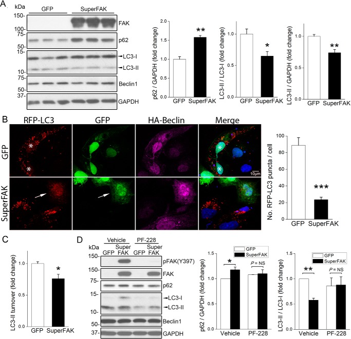
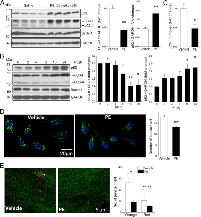
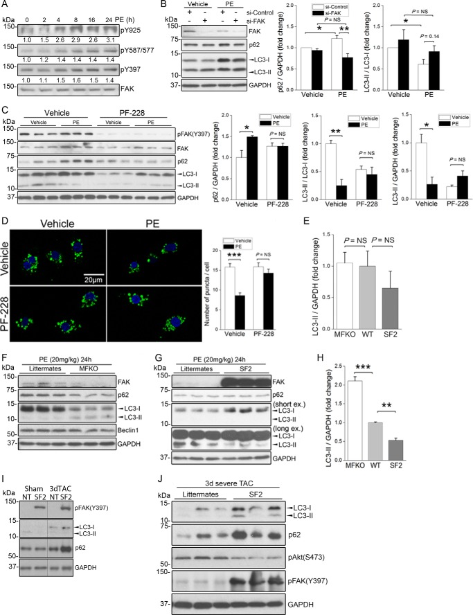
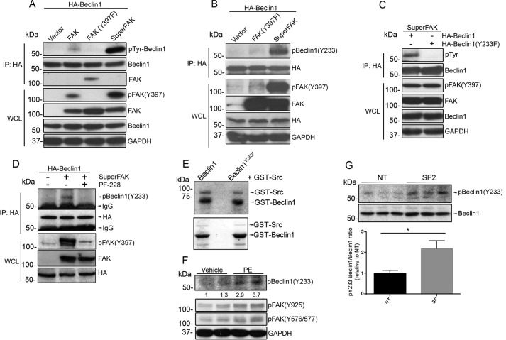
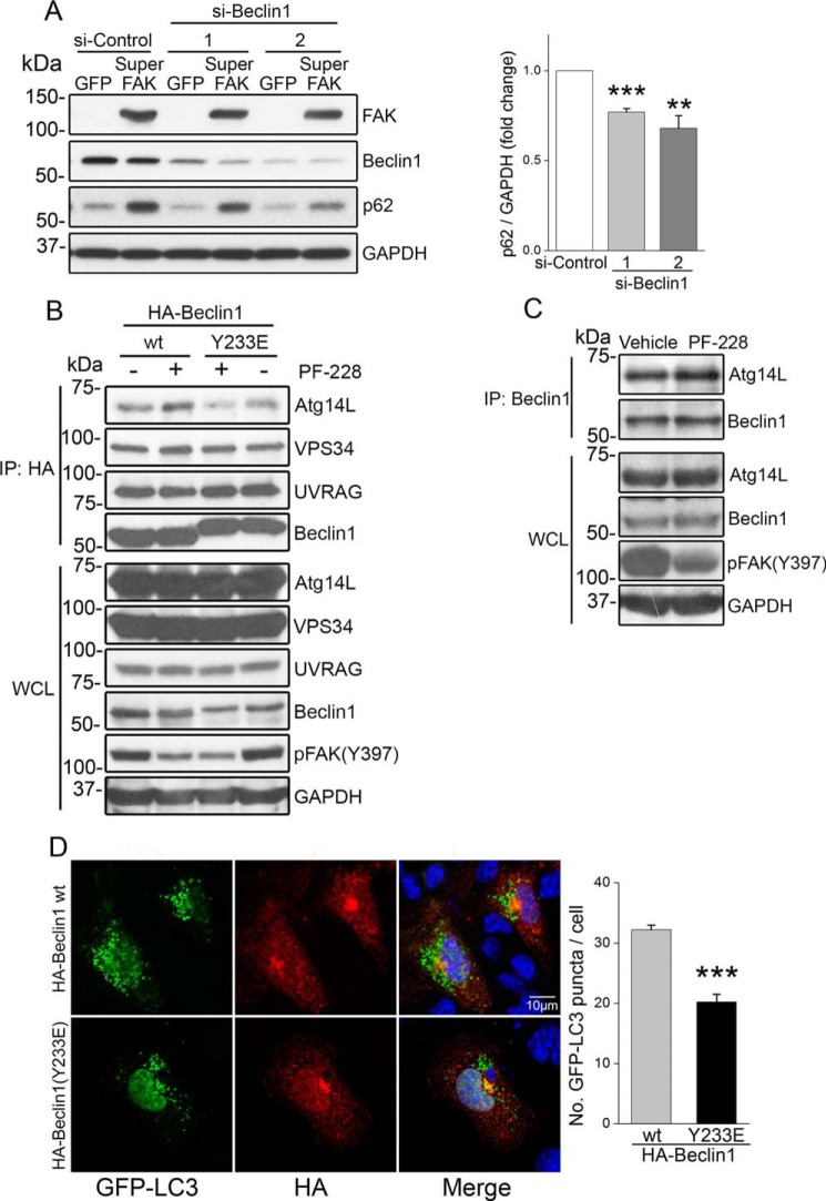
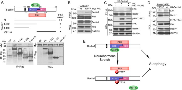
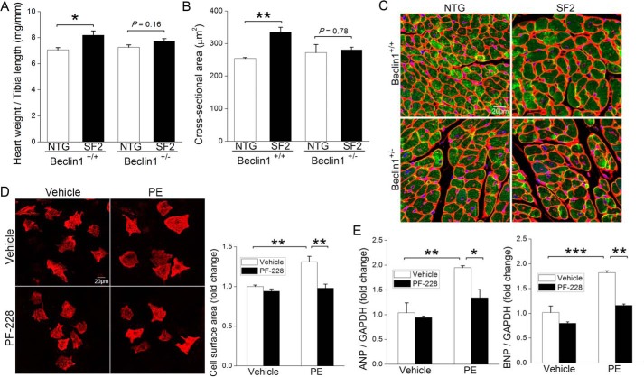
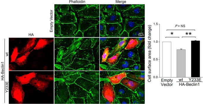
Autophagy is an evolutionarily conserved intracellular degradation/recycling system that is essential for cellular homeostasis but is dysregulated in a number of diseases, including myocardial hypertrophy. Although it is clear that limiting or accelerating autophagic flux can result in pathological cardiac remodeling, the physiological signaling pathways that fine-tune cardiac autophagy are poorly understood. Herein, we demonstrated that stimulation of cardiomyocytes with phenylephrine (PE), a well known hypertrophic agonist, suppresses autophagy and that activation of focal adhesion kinase (FAK) is necessary for PE-stimulated autophagy suppression and subsequent initiation of hypertrophic growth. Mechanistically, we showed that FAK phosphorylates Beclin1, a core autophagy protein, on Tyr-233 and that this post-translational modification limits Beclin1 association with Atg14L and reduces Beclin1-dependent autophagosome formation. Remarkably, although ectopic expression of wild-type Beclin1 promoted cardiomyocyte atrophy, expression of a Y233E phosphomimetic variant of Beclin1 failed to affect cardiomyocyte size. Moreover, genetic depletion of Beclin1 attenuated PE-mediated/FAK-dependent initiation of myocyte hypertrophy in vivo Collectively, these findings identify FAK as a novel negative regulator of Beclin1-mediated autophagy and indicate that this pathway can facilitate the promotion of compensatory hypertrophic growth. This novel mechanism to limit Beclin1 activity has important implications for treating a variety of pathologies associated with altered autophagic flux.

Keywords: Beclin-1 (BECN1); PTK2 protein tyrosine kinase 2 (PTK2) (focal adhesion kinase) (FAK); atrophy; autophagy; cardiac hypertrophy; muscle atrophy.


Conflict of interest statement

The authors declare that they have no conflicts of interest with the contents of this article

Figures

FIGURE 1.
FIGURE 1.
Activation of FAK limits cardiomyocyte autophagy. A, NRCMs were infected with GFP or SuperFAK adenoviruses for 72 h, and cell lysates were blotted with indicated antibodies with GAPDH as a loading control. Quantification shown on right, n = 3; *, p < 0.05; **, p < 0.01 versus GFP. B, HL-1 cardiomyocytes were co-transfected with RFP-LC3 or HA-Beclin1 for 30 h, followed by infection with GFP or SuperFAK adenoviruses for 18 h and starvation for 4 h in the presence of CQ. Cells were then fixed with 4% paraformaldehyde and stained to detect HA (purple) and nuclei (DAPI, blue). GFP- (asterisk) or SuperFAK-infected cells appear green. Overexpression of SuperFAK reduced the number of RFP-LC3 puncta (autophagosomes, arrow). ***, p < 0.001 versus GFP. Quantification represents mean ± S.E. of three independent experiments (at least 50 cells positive for RFP-LC3, GFP, and HA-Beclin1 were analyzed in each group). Scale bar, 10 μm. C, NRCMs were infected with GFP or SuperFAK adenoviruses for 48 h. The lysosomal inhibitor CQ (10 μm) was added to half of the samples to assess autophagic flux. LC3-II turnover is defined as the amount of LC3-II delivered to lysosomes for degradation within a certain period of time and calculated by subtracting LC3-II band intensity in the presence of CQ with that in the absence of CQ. *, p < 0.05 versus GFP. D, NRCMs were infected with GFP or SuperFAK adenoviruses for 48 h with or without the potent and selective FAK inhibitor PF-228 in the last 24 h. *, p < 0.05; **, p < 0.01. NS, not significant. Data are mean ± S.E. of three independent experiments. All data were analyzed using two-tailed Student's t tests.
FIGURE 2.
FIGURE 2.
Activation of α-adrenergic signaling suppresses cardiac autophagy. A, C57BL/6 mice received a single injection of PE (20 mg/kg, s.c.) or saline control (n = 4 in each group). Hearts were harvested. and protein lysates were blotted using indicated antibodies with GAPDH as a loading control. B, NRCMs were treated with PE (50 μm) for various times from 2 to 24 h. Cell lysates were blotted with indicated antibodies. Quantification of p62 and LC3-II/LC3-I ratio and LC3-II levels are shown on right in A and B. *, p < 0.05; **, p < 0.01 versus non-treated control. Results are mean ± S.E. of three independent experiments. C, NRCMs were incubated with PE (50 μm) for 24 h with or without CQ (10 μm) in the last 4 h of treatment. LC3-II turnover was calculated by subtracting LC3-II band intensity in the presence of CQ with that in the absence of CQ. *, p < 0.05 versus vehicle. D, NRCMs were treated with PE (50 μm) or vehicle for 4 h in the presence of CQ (10 μm). Cells were subsequently stained with the CYTO-ID® autophagy detection kit was used to visualize autophagic vacuoles (green puncta). Treatment with PE significantly reduced puncta formation. *, p < 0.05 versus vehicle. At least 50 cells were examined per sample. Data are mean ± S.E. of three independent experiments. Scale bar, 20 μm. **, p < 0.01. E, CAG-RFP-EGFP-LC3 mice were transferred to food-free cages and received a single injection of PE (20 mg/kg, s.c.) or saline control (n = 3 in each group), and hearts were collected at 24 h. The lysosomal acidification inhibitor bafilomycin A1 was injected 2 h before euthanization. Autophagosomes appear orange, and autolysosomes appear red. Number of puncta was quantified from at least five different high power fields. *, p < 0.05. NS, not significant. Data were analyzed using two-tailed Student's t tests.
FIGURE 3.
FIGURE 3.
α-Adrenergic-dependent autophagy suppression requires FAK. A, NRCMs were treated with PE (50 μm) for various times from 2 to 24 h. Cell lysates were blotted with indicated antibodies. Identical lysates were used in Fig. 2B. Results are representative of three independent experiments. Quantification of FAK phosphorylation/total FAK is indicated (relative to the 0 time point) for three separate experiments. B, NRCMs were transfected with control or FAK siRNA (25 nm) for 72 h prior to treatment with PE (50 μm) for 24 h. Cell lysates were blotted with indicated antibodies (right panel). Data were quantified by densitometry and graphed as mean ± S.E. of three independent experiments; *, p < 0.05; **, p < 0.01. C, NRCMs were pre-treated with the pharmacological FAK inhibitor PF-228 (1 μm) for 1 h prior to the addition of PE (50 μm) for 24 h. Pre-treatment with FAK inhibitor blocked PE-mediated autophagic suppression (right panel). Data were quantified by densitometry and graphed as mean ± S.E. of three independent experiments; *, p < 0.05; **, p < 0.01. D, NRCMs were pre-treated with FAK inhibitor PF-228 (1 μm) for 1 h prior to incubation with PE (50 μm) for 4 h in the presence of CQ (10 μm). Autophagic vesicles were detected by staining with the CYTO-ID® autophagy detection kit. ***, p < 0.001. Scale bar, 20 μm. E, quantification of LC3-II levels in basal WT, MFKO myocyte-restricted FAK knock-out, and SF2 hearts (NS, not significant; n = 3–5 per group). F–H, MFKO, SF2 mice and WT littermates received a single injection of PE (20 mg/kg, s.c.). Hearts were harvested at 24 h, and protein lysates were blotted using indicated antibodies. Blots are representative of 3–5 hearts per group. H, quantification of LC3-II levels in PE-treated WT, MFKO, and SF2 hearts (**, p < 0.01; ***, p < 0.001). I and J, SF2 mice were subjected to moderate (I) or severe (J) TAC. Hearts were isolated at day 3, and protein lysates were blotted with indicated antibodies. Data are representative of three hearts per group.
FIGURE 4.
FIGURE 4.
FAK induces Beclin1 phosphorylation on Tyr-233. A, COS cells were co-transfected with HA-Beclin1 and wild-type FAK, non-phosphorylatable FAK mutant (Y397F), or SuperFAK. At 24 h, cell lysates were subjected to immunoprecipitation (IP) with anti-HA followed by Western blotting with indicated antibodies, including an anti-phosphotyrosine antibody (Tyr(P), 4G10). WCL, whole cell lysate. B, protein lysates from COS cells co-transfected with HA-Beclin1 and non-phosphorylatable FAK mutant (Y397F) or SuperFAK were immunoprecipitated with an anti-HA antibody followed by Western blotting analysis with anti-phospho-Beclin1 (Tyr-233). C, COS cells were co-transfected with SuperFAK and HA-Beclin1 or a non-phosphorylatable Beclin1 mutant (Y233F). Cell lysates were immunoprecipitated with an anti-HA antibody followed by Western blotting analysis with an anti-phosphotyrosine antibody (Tyr(P), 4G10). D, COS cells were co-transfected with SuperFAK and HA-Beclin1 prior to treatment with FAK inhibitor PF-228 for 16 h. Cell lysates were immunoprecipitated with an anti-HA antibody followed by Western blotting analysis with anti-phospho-Beclin1 (Tyr-233). E, in vitro Src kinase assay. Purified GST-Beclin1 and GST-Beclin1-Y233F MAPK were incubated with GST-Src and [γ-32P]ATP. Samples were analyzed by SDS-PAGE, and the gel was dried and exposed to Kodak XAR film for 2 h, followed by Coomassie Blue staining. Gel is representative of four separate experiments. F, NRCMs were treated with PE (50 μm) for 8 h. Duplicate cell lysates were blotted with indicated antibodies. G, cardiac-specific SuperFAK transgenic (SF2) mice and littermates received a single injection of PE (20 mg/kg, s.c.). Hearts were harvested at 24 h, and protein lysates were immunoprecipitated with Beclin1 antibody followed by immunoblotting using indicated antibodies. Blots represent three hearts per group, and data shown are mean ± S.E.; p < 0.05.
FIGURE 5.
FIGURE 5.
Phosphorylation of Tyr-233 by FAK limits Beclin1-dependent autophagy. A, Beclin1 is required for SuperFAK-mediated autophagy suppression. NRCMs were infected with GFP or SuperFAK adenoviruses and transfected with control or Beclin1 siRNAs for 48 h prior to treatment with PE (50 μm) for 24 h. Cell lysates were blotted with indicated antibodies with GAPDH as a loading control. Quantification of mean ± S.E. of three independent experiments revealed that knockdown of Beclin1 reduced SuperFAK-induced increase in p62 protein levels when compared with si-Control (right panel). **, p < 0.01; ***, p < 0.001. B, FAK activity and FAK-dependent phosphorylation of Beclin1 alters the Beclin1 interactome. HL-1 cells were transfected with HA-Beclin1 or a phosphomimetic Beclin1 mutant (Y233E) for 48 h prior to starvation in the presence of FAK inhibitor PF-228 (1 μm) or vehicle control for 22 h. Lysates were immunoprecipitated with anti-HA and subjected to Western blotting with indicated antibodies. C, co-immunoprecipitation of endogenous Beclin1 with Atg14L in starved HL-1 cardiomyocytes treated with PF-228 (1 μm) or vehicle control for 22 h. D, Beclin Y233E phosphomimetic has limited capacity to induce autophagy. HL-1 cells were co-transfected with GFP-LC3 and HA-Beclin1 or a phosphomimetic Beclin1 variant (Y233E) for 48 h prior to treatment with starvation in the presence of FAK inhibitor PF-228 (1 μm) and CQ (10 μm) for 4 h. Cells were fixed and stained for GFP (green), HA (red), and nuclei (DAPI, blue). Beclin1(Y233E)-expressing cells exhibited less GFP-LC3 puncta (autophagosomes) than wild-type Beclin1-expressing cells. ***, p < 0.001 versus wild-type Beclin1. Results are mean ± S.E. of three independent experiments (>50 cells were analyzed each group). Scale bar, 10 μm. All data were analyzed using two-tailed Student's t tests.
FIGURE 6.
FIGURE 6.
FAK associates with Beclin1 in an activation-specific fashion. A, top, schematic representation of three functional domains of Beclin1 (the N-terminal Bcl-2 homology 3 domain (BH3), the central coiled-coil domain (CCD), and the C-terminal ECD), and FLAG-tagged (FL) Beclin1 deletion constructs. Bottom, FAK interaction sites on Beclin1 were mapped by co-immunoprecipitation of Beclin1 with FAK in COS cells co-transfected with Myc-FAK and indicated FLAG-Beclin1 constructs. B, immunoprecipitation of Beclin1 with FAK in COS cells co-transfected with HA-Beclin1 and wild-type FAK or the non-phosphorylatable FAK mutant (Y397F). C, immunoprecipitation of Beclin1 with FAK in COS cells co-transfected with HA-Beclin1 and wild-type FAK and incubated ± FAK inhibitor 14 (50 μm for 1 h) prior to lysis. D, immunoprecipitation of Beclin1 with FAK in COS cells co-transfected with FAK(Y397F) and wild-type Beclin1 or the non-phosphorylatable Beclin1 variant (Y233F). E, model depicting the effect of FAK-mediated Beclin1 phosphorylation on autophagy initiation. Under conditions of nutrient starvation, Beclin1 forms a complex with Atg14L to promote autophagy. In response to neurohormone stimulation or mechanical stretch, FAK-mediated phosphorylation of Tyr-233 Beclin1 disrupts the Beclin1-Atg14L interaction and suppresses autophagy.
FIGURE 7.
FIGURE 7.
FAK-mediated cardiomyocyte hypertrophy requires Beclin1 and FAK activity. A–C, cardiac-specific SuperFAK transgenic (SF2) or non-transgenic (NTG) mice on a Beclin1+/+ or Beclin1+/− background received a single injection of PE (20 mg/kg, s.c.), and hearts were harvested 24 h following treatment. A, heart weight normalized to tibia length. B and C, myocyte cross-sectional area as assessed by staining with wheat germ agglutinin (red), cardiac troponin T (green), and DAPI (blue). At least 200 cells from five different fields were analyzed in each heart. *, p < 0.05; **, p < 0.01. Data are mean ± S.E. from 3 to 4 animals per group. D–F, pre-treatment with FAK inhibitor abrogated PE-induced cardiomyocyte hypertrophy. NRCMs were treated with PE (50 μm) with or without FAK inhibitor PF-228 (1 μm) for 48 h. D, cardiac myocytes were labeled with anti-cardiac troponin T (red), and cell surface area was quantified using ImageJ software (National Institutes of Health). **, p < 0.01. Data are mean ± S.E. of four independent experiments (>200 cells were analyzed each group). Scale bar, 20 μm. E, quantitative RT-PCR analysis of hypertrophy markers ANP (left) and BNP (right). *, p < 0.05; **, p < 0.01; ***, p < 0.001. Data are mean ± S.E. of three independent experiments and were analyzed using two-tailed Student's t tests. ANP, atrial natriuretic peptide; BNP, brain natriuretic peptide.
FIGURE 8.
FIGURE 8.
Beclin1 but not Y233E Beclin1 induces cardiomyocyte atrophy. HL-1 cells were transfected with HA-Beclin1 WT, HA-Beclin1 Y233E variant, or empty vector for 48 h. Cells were then fixed in 2% paraformaldehyde and stained with HA (red), phalloidin (green), and DAPI (blue). HL-1 cell size was measured using ImageJ software. Results are mean ± S.E. of three independent experiments (>50 cells were analyzed each group). Scale bar, 10 μm. NS, not significant. Data were analyzed by one-way analysis of variance with Dunnett's post hoc test.

References

    1. Wesselborg S., and Stork B. (2015) Autophagy signal transduction by atg proteins: From hierarchies to networks. Cell. Mol. Life Sci. 72, 4721–4757 - PMC - PubMed
    1. Rothermel B. A., and Hill J. A. (2008) Autophagy in load-induced heart disease. Circ. Res. 103, 1363–1369 - PMC - PubMed
    1. Gustafsson A. B., and Gottlieb R. A. (2009) Autophagy in ischemic heart disease. Circ. Res. 104, 150–158 - PMC - PubMed
    1. Nakai A., Yamaguchi O., Takeda T., Higuchi Y., Hikoso S., Taniike M., Omiya S., Mizote I., Matsumura Y., Asahi M., Nishida K., Hori M., Mizushima N., and Otsu K. (2007) The role of autophagy in cardiomyocytes in the basal state and in response to hemodynamic stress. Nat. Med. 13, 619–624 - PubMed
    1. Kanamori H., Takemura G., Goto K., Maruyama R., Tsujimoto A., Ogino A., Takeyama T., Kawaguchi T., Watanabe T., Fujiwara T., Fujiwara H., Seishima M., and Minatoguchi S. (2011) The role of autophagy emerging in postinfarction cardiac remodelling. Cardiovasc. Res. 91, 330–339 - PubMed

Publication types

Substances