. 2016 Dec 7:11:6499-6506.
doi: 10.2147/IJN.S41371. eCollection 2016.

Reduced Staphylococcus aureus biofilm formation in the presence of chitosan-coated iron oxide nanoparticles

Affiliations

Reduced Staphylococcus aureus biofilm formation in the presence of chitosan-coated iron oxide nanoparticles

Si-Feng Shi et al. Int J Nanomedicine. .

Abstract

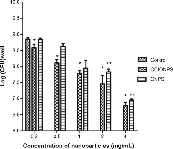
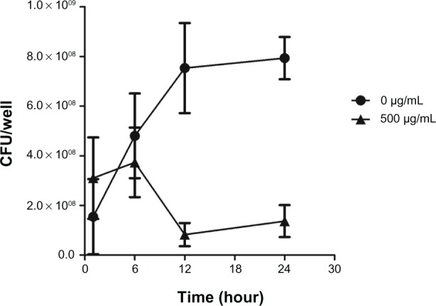
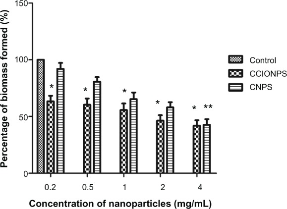
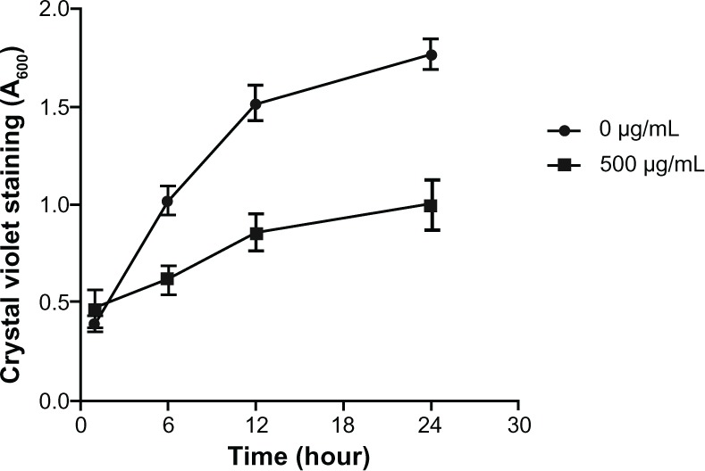
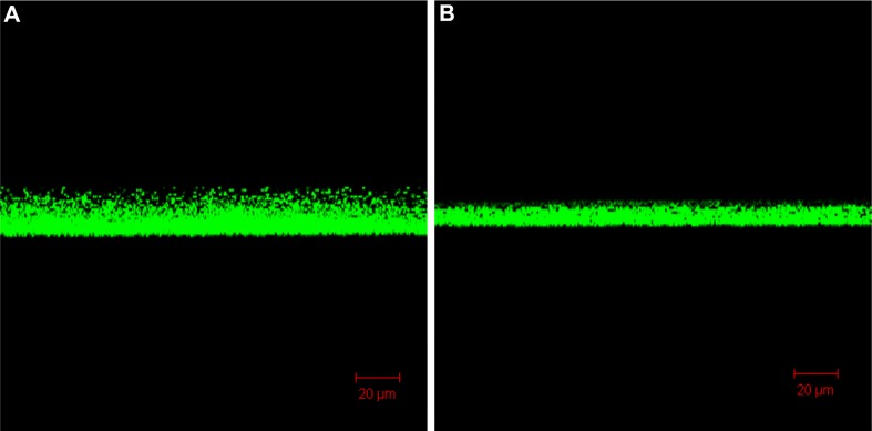
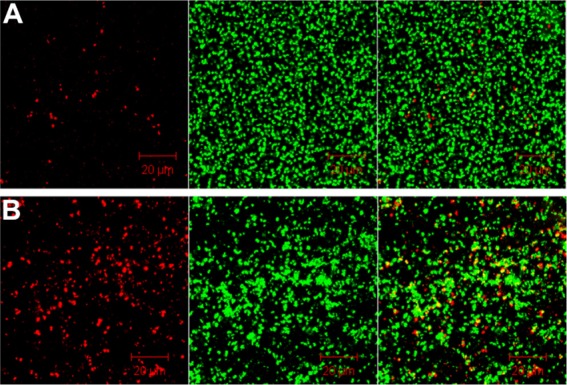
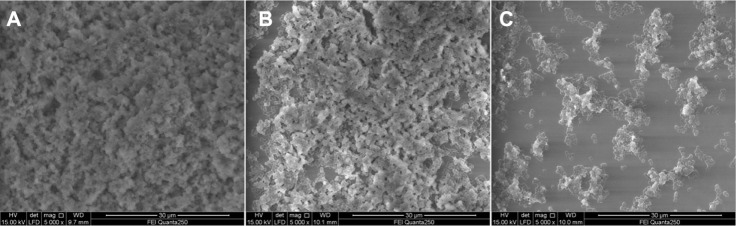
Staphylococcus aureus can adhere to most foreign materials and form biofilm on the surface of medical devices. Biofilm infections are difficult to resolve. The goal of this in vitro study was to explore the use of chitosan-coated nanoparticles to prevent biofilm formation. For this purpose, S. aureus was seeded in 96-well plates to incubate with chitosan-coated iron oxide nanoparticles in order to study the efficiency of biofilm formation inhibition. The biofilm bacteria count was determined using the spread plate method; biomass formation was measured using the crystal violet staining method. Confocal laser scanning microscopy and scanning electron microscopy were used to study the biofilm formation. The results showed decreased viable bacteria numbers and biomass formation when incubated with chitosan-coated iron oxide nanoparticles at all test concentrations. Confocal laser scanning microscopy showed increased dead bacteria and thinner biofilm when incubated with nanoparticles at a concentration of 500 µg/mL. Scanning electron microscopy revealed that chitosan-coated iron oxide nanoparticles inhibited biofilm formation in polystyrene plates. Future studies should be performed to study these nanoparticles for anti-infective use.

Keywords: Staphylococcus aureus; biofilm; chitosan-coated iron oxide nanoparticles.


Conflict of interest statement

The authors report no conflicts of interest in this work.

Figures

Figure 1
Figure 1
Effects of different concentrations of nanoparticles on biofilm bacterial growth at after 24 hours of incubation. Notes: The results are expressed as mean ± SD (n = 3). *P < 0.05 compared with control sample; **P < 0.05 compared with control sample (blank). Abbreviations: CCIONPS, chitosan-coated iron oxide nanoparticles; CFU, colony-forming unit; CNPS, chitosan nanoparticles; SD, standard deviation.
Figure 2
Figure 2
Effects of chitosan-coated iron oxide nanoparticles on biofilm bacterial growth in the first 24 hours at the concentration of 500 µg/mL. Note: The results are expressed as mean ± SD (n = 3). Abbreviations: CFU, colony-forming unit; SD, standard deviation.
Figure 3
Figure 3
Relative biomass formed after 24 hours of incubation with nanoparticles at different concentrations. Notes: The results are expressed as mean ± SD (n = 3). *P < 0.05 compared with control sample; **P < 0.05 compared with samples at concentrations of 200 µg/mL and 500 µg/mL. Abbreviations: CCIONPS, chitosan-coated iron oxide nanoparticles; CNPS, chitosan nanoparticles; SD, standard deviation.
Figure 4
Figure 4
Biomass formed in the first 24 hours of incubation with chitosan-coated iron oxide nanoparticles at a concentration of 500 µg/mL. Note: The results are expressed as mean ± SD (n = 3). Abbreviation: SD, standard deviation.
Figure 5
Figure 5
Confocal images show the biofilm thickness after 48 hours of incubation. (A) In the absence of nanoparticles; (B) incubated with chitosan-coated iron oxide nanoparticles at a concentration of 500 µg/mL.
Figure 6
Figure 6
Confocal images show dead bacteria after 48 hours of incubation. (A) In the absence of nanoparticles; (B) incubated with chitosan-coated iron oxide nanoparticles at a concentration of 500 µg/mL. Note: Dead cells (left), living cells (middle), and overlapping images (right).
Figure 7
Figure 7
SEM images showing biofilms of the bacteria established on the surface of polystyrene plates. (A) In the absence of nanoparticles; (B) incubated with chitosan-coated iron oxide nanoparticles at 500 µg/mL; (C) incubated with chitosan-coated iron oxide nanoparticles at 4 mg/mL. Abbreviation: SEM, scanning electron microscopy.

References

    1. Zoubos AB, Galanakos SP, Soucacos PN. Orthopedics and biofilm – what do we know? A review. Med Sci Monit. 2012;18(6):RA89–RA96. - PMC - PubMed
    1. Subramani K, Jung RE, Molenberg A, Hammerle CH. Biofilm on dental implants: a review of the literature. Int J Oral Maxillofac Implants. 2009;24(4):616–626. - PubMed
    1. Shi S, Zhang X. Interaction of Staphylococcus aureus with osteoblasts (Review) Exp Ther Med. 2012;3(3):367–370. - PMC - PubMed
    1. Urquhart DM, Hanna FS, Brennan SL, et al. Incidence and risk factors for deep surgical site infection after primary total hip arthroplasty: a systematic review. J Arthroplasty. 2010;25(8):1216–1222. e1–e3. - PubMed
    1. Lellouche J, Kahana E, Elias S, Gedanken A, Banin E. Antibiofilm activity of nanosized magnesium fluoride. Biomaterials. 2009;30(30):5969–5978. - PubMed

MeSH terms