Review
. 2016 Dec;50(4):275-283.
doi: 10.1007/s13139-015-0380-y. Epub 2015 Oct 22.

Stem Cell Monitoring with a Direct or Indirect Labeling Method

Affiliations
Review

Stem Cell Monitoring with a Direct or Indirect Labeling Method

Min Hwan Kim et al. Nucl Med Mol Imaging. 2016 Dec.

Abstract

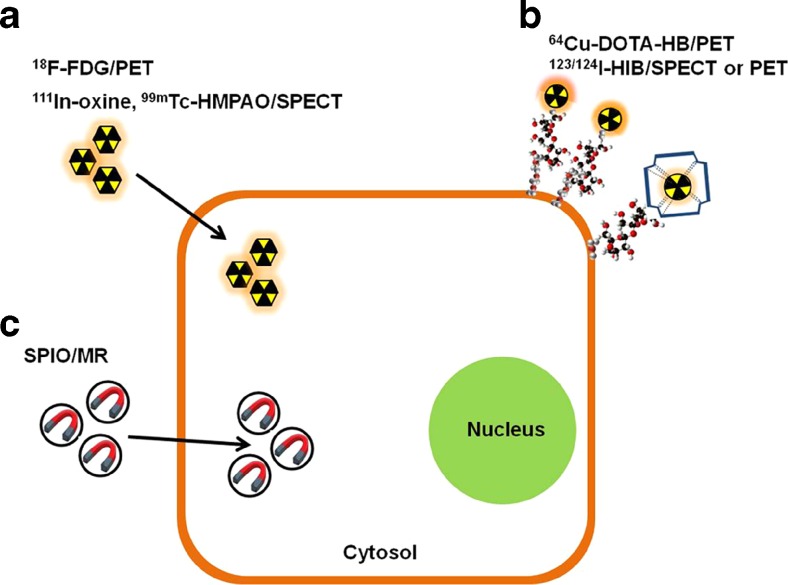
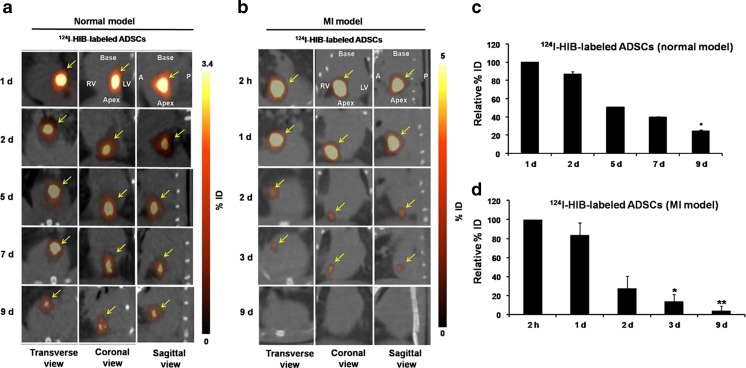
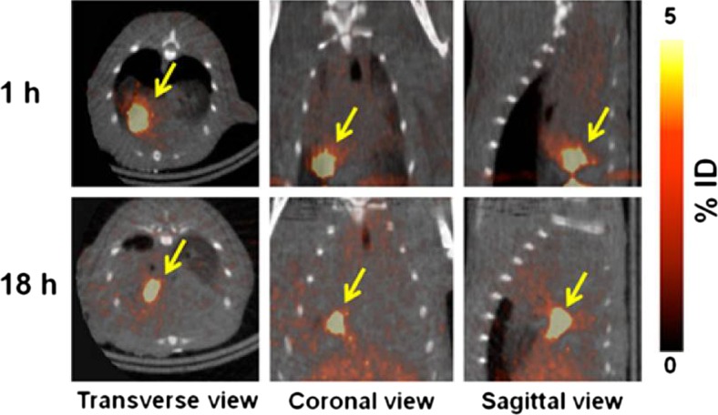
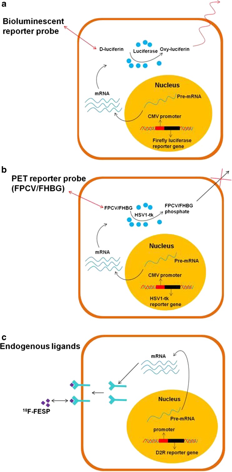
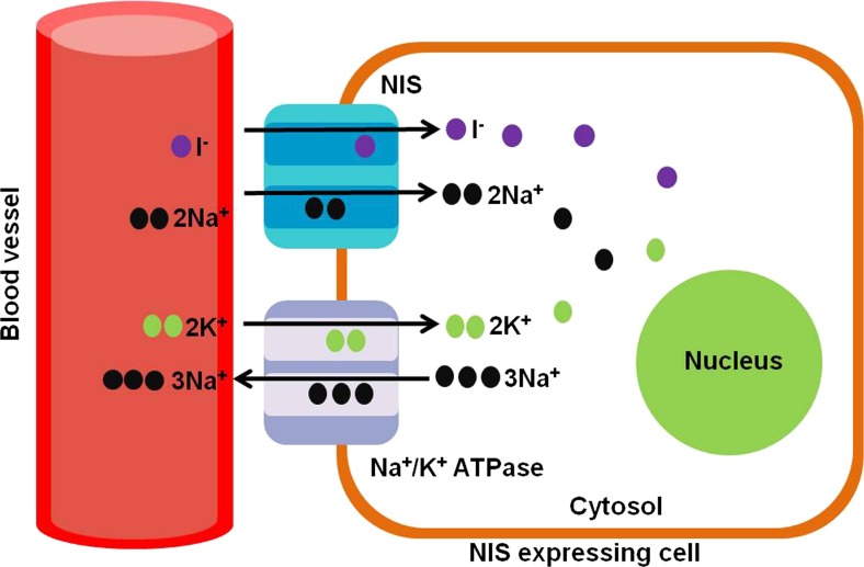
The molecular imaging techniques allow monitoring of the transplanted cells in the same individuals over time, from early localization to the survival, migration, and differentiation. Generally, there are two methods of stem cell labeling: direct and indirect labeling methods. The direct labeling method introduces a labeling agent into the cell, which is stably incorporated or attached to the cells prior to transplantation. Direct labeling of cells with radionuclides is a simple method with relatively fewer adverse events related to genetic responses. However, it can only allow short-term distribution of transplanted cells because of the decreasing imaging signal with radiodecay, according to the physical half-lives, or the signal becomes more diffuse with cell division and dispersion. The indirect labeling method is based on the expression of a reporter gene transduced into the cell before transplantation, which is then visualized upon the injection of an appropriate probe or substrate. In this review, various imaging strategies to monitor the survival and behavior change of transplanted stem cells are covered. Taking these new approaches together, the direct and indirect labeling methods may provide new insights on the roles of in vivo stem cell monitoring, from bench to bedside.

Keywords: Direct labeling method; Indirect labeling method; Radionuclide; Stem cell tracking.


Conflict of interest statement

Min Hwan Kim, Yong Jin Lee, and Joo Hyun Kang declare that they have no conflict of interest, etc. Ethical Statement The care, maintenance, and treatment of animals in these studies followed protocols approved by the Institutional Animal Care and Use Committee of the Korea Institute of Radiological and Medical Sciences (KIRAMS).

Figures

Fig. 1
Fig. 1
Direct cell labeling methods. In a direct labeling strategy, various labeling agents are for radionuclide imaging (a, b) or MRI (c) and are introduced into the inner part of stem cells (a, c, e.g., 18F-FDG, 111In-oxine, 99mTc-HMPAO, or SPIO) or anchored to the cell membrane (b, e.g., 64Cu-DOTA-HB, 123/124I-HIB). To track the transplanted stem cells, first, the direct-labeled stem cells are transplanted to the target tissue/organ. Then, noninvasive imaging including MRI or radionuclide imaging using PET or SPECT is performed at the target site for detection of transplanted stem cells
Fig. 2
Fig. 2
Small-animal PET/CT imaging of transplanted 124I-HIB-labeled ADSCs in the normal and MI model and longitudinal quantification of the relative percent of the injected dose (%ID). a, b Small-animal PET/CT images of transplanted 124I-HIB-labeled ADSCs in the normal (a) and MI model (b) for 9 days. c, d Longitudinal quantification of the relative %ID of transplanted 124I-HIB-labeled ADSCs in the normal (c, * P < 0.05 vs. day 1) and MI model (d, * P < 0.05, ** P < 0.001 vs. 2 h) for 9 days. Arrows indicate the injection site of 124I-HIB-labeled ADSCs. A: anterior, P: posterior, RV: right ventricle, LV: left ventricle
Fig. 3
Fig. 3
Small-animal PET/CT image of intra-myocardially injected-labeled ADSCs (1.8−3.7 MBq, 100 μl) in normal heart for 18 h. Arrow indicates the site of 64Cu-DOTA-HB-labeled ADSC injection
Fig. 4
Fig. 4
Various types of reporter gene/reporter probe strategies. a Enzyme-based bioluminescence imaging. D-luciferin is a substrate molecular probe that is acted upon by the enzyme firefly luciferase (fLuc) to result in bioluminescence via a chemiluminescent reaction under physiological conditions only within living cells expressing the fLuc gene. b Enzyme-based PET imaging. 8-18F-fluoropenciclovir (18F-FPCV) or other acycloguanosines are substrate molecular probes phosphorylated by the herpes simplex virus type 1 thymidine kinase (HSV1-tk) enzyme to result in the intracellular trapping of the probe in cells expressing the HSV1-tk gene. c Receptor-based PET imaging. 3-(2′-18F-fluoroethyl)spiperone (18F-FESP) is a ligand molecular probe interacting with the dopamine receptor D2 (D2R) to result in trapping of the probe on/in cells expressing the D2R gene
Fig. 5
Fig. 5
Schematic presentation of NIS function. NIS transports two sodium ions and one iodide ion into the cytoplasm together. The electrochemical sodium gradient generated by the Na+/K+ ATPase pump provides energy for this transfer
Fig. 6
Fig. 6
In vivo cell fate in a canine MI model. a Representative transaxial 99mTcO4 SPECT images of the saline vehicle control group and experimental group. When compared to the saline vehicle control group, the experimental group showed distinguishable 99mTc accumulation until day 9, which reflects the viability of injected stem cells in the left ventricular wall. b Time course of myocardial accumulation of 99mTc-TcO4 counted from serial 99mTc-TcO4 SPECT images in the saline vehicle control group and experimental group
Fig. 7
Fig. 7
Expression of cardiac-specific genes and stem cell-specific markers according to cardiomyogenic differentiation of BMSCs. a Expression of cardiac-specific genes (α-MHC, MLC-2V) and a stem cell-specific marker (Sca-1) in cardiomyogenic differentiated BMSCs treated with ATRA. b Relative quantification of gene expression normalized against GAPDH. c Radioiodine (125I) uptake of cultured BMSCs with or without ATRA in vitro. Cardiomyogenic differentiated BMSCs treated with ATRA showed higher 125I uptake than nontreated BMSCs

References

    1. Hsiao LC, Carr C, Chang KC, Lin SZ, Clarke K. Stem cell-based therapy for ischemic heart disease. Cell Transplant. 2013;22:663–75. doi: 10.3727/096368912X655109. - DOI - PubMed
    1. Jazi SM, Esfahani MH, Fesharaki M, Moulavi F, Gharipour M. Initial clinical outcomes of intracoronary infusion of autologous progenitor cells in patients with acute myocardial infarction. ARYA Atheroscler. 2012;7:162–7. - PMC - PubMed
    1. Welt FG, Losordo DW. Cell therapy for acute myocardial infarction: curb your enthusiasm? Circulation. 2006;113:1272–4. doi: 10.1161/CIRCULATIONAHA.105.613034. - DOI - PubMed
    1. Al-Nbaheen M, Vishnubalaji R, Ali D, Bouslimi A, Al-Jassir F, Megges M, et al. Human stromal (mesenchymal) stem cells from bone marrow, adipose tissue and skin exhibit differences in molecular phenotype and differentiation potential. Stem Cell Rev. 2013;9:32–43. doi: 10.1007/s12015-012-9365-8. - DOI - PMC - PubMed
    1. De Coppi P, Bartsch G, Jr, Siddiqui MM, Xu T, Santos CC, Perin L, et al. Isolation of amniotic stem cell lines with potential for therapy. Nat Biotechnol. 2007;25:100–6. doi: 10.1038/nbt1274. - DOI - PubMed