. 2016 Dec 13:4:e2682.
doi: 10.7717/peerj.2682. eCollection 2016.

Malagasy Conostigmus (Hymenoptera: Ceraphronoidea) and the secret of scutes

Affiliations

Malagasy Conostigmus (Hymenoptera: Ceraphronoidea) and the secret of scutes

István Mikó et al. PeerJ. .

Abstract

We revise the genus Conostigmus Dahlbom 1858 occurring in Madagascar, based on data from more specimens than were examined for the latest world revision of the genus. Our results yield new information about intraspecific variability and the nature of the atypical latitudinal diversity gradient (LDG) observed in Ceraphronoidea. We also investigate cellular processes that underlie body size polyphenism, by utilizing the correspondence between epidermal cells and scutes, polygonal units of leather-like microsculpture. Our results reveal that body size polyphenism in Megaspilidae is most likely related to cell number and not cell size variation, and that cell size differs between epithelial fields of the head and that of the mesosoma. Three species, Conostigmus ballescoracas Dessart, 1997, C. babaiax Dessart, 1996 and C. longulus Dessart, 1997, are redescribed. Females of C. longulus are described for the first time, as are nine new species: C. bucephalus Mikó and Trietsch sp. nov., C. clavatus Mikó and Trietsch sp. nov., C. fianarantsoaensis Mikó and Trietsch sp. nov., C. lucidus Mikó and Trietsch sp. nov., C. macrocupula, Mikó and Trietsch sp. nov., C. madagascariensis Mikó and Trietsch sp. nov., C. missyhazenae Mikó and Trietsch sp. nov., C. pseudobabaiax Mikó and Trietsch sp. nov., and C. toliaraensis Mikó and Trietsch sp. nov. A fully illustrated identification key for Malagasy Conostigmus species and a Web Ontology Language (OWL) representation of the taxonomic treatment, including specimen data, nomenclature, and phenotype descriptions, in both natural and formal languages, are provided.

Keywords: CLSM; Imaginal disks; LDG; Male genitalia; Microscopy; Morphology; Phenotypic plasticity; Taxonomy.


Conflict of interest statement

The authors declare that they have no competing interests.

Figures

Figure 1
Figure 1. CLSM volume rendered micrographs showing the skeletomuscular system of the male genitalia of Conostigmus longulus Dessart, 1997.
(A) Excitation wavelength = 631 nm, emission wavelength = 647 nm. (B) Excitation wavelength = 499 nm, emission wavelength = 520 nm. Abbreviations: pss, parossiculus; gs, gonostipes; hrp, harpe; gs-hrp, proximal gonostyle/volsella complex-harpal muscle; imvl, medial gonostyle volsella complex-volsellal muscle.
Figure 2
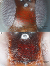
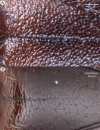
Figure 2. Brightfield image showing the median region of the cranium of Conostigmus longulus Dessart, 1997, anterior view.
The measured scute lengths and values and borders of measured rectangle are annotated in red color. Size of rectangular area = 9,636 μm2.
Figure 3
Figure 3. Relationship between body size and number of cells as linear regression.
Interorbital space length (IOS), measured in μm, stands as a proxy for body size. Number of cells refers to the number of scutes/cells of a standard sized rectangular area. The size of these cells is not correlated to significant body size differences (Fig. S4).
Figure 4
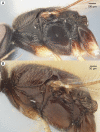
Figure 4. Brightfield image showing the lateral habitus of Conostigmus babaiax Dessart, 1997.
Figure 5
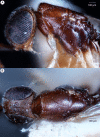
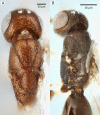
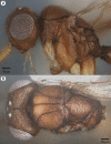
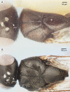
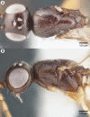
Figure 5. Brightfield image showing the head and mesosoma of Conostigmus babaiax Dessart, 1997.
(A) Lateral view. (B) Dorsal view.
Figure 6
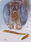
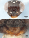
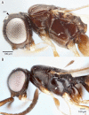
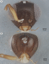
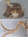
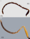
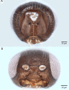
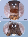
Figure 6. Brightfield image showing the head and female antenna of Conostigmus babaiax Dessart, 1997.
(A) Head, anterior view. (B) Female antenna.
Figure 7
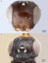
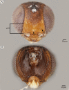
Figure 7. Brightfield image showing the head of Conostigmus ballescoracas Dessart, 1997.
(A) Anterior view. (B) Dorsal view (ar, randomly sized areolae around setal pits; poc, preoccipital carina).
Figure 8
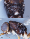
Figure 8. Brightfield image of Conostigmus ballescoracas Dessart, 1997.
(A) Head and mesosoma lateral view. (B) Female antenna lateral view (poc, preoccipital carina; app, anteromedian projection of the metanoto-propodeo-metapecto-mesopectal complex; usc, u-shaped carina surrounding posteriorly and laterally the disc of the mesoscutellum).
Figure 9
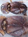
Figure 9. Brightfield image of Conostigmus ballescoracas Dessart, 1997.
(A) Mesosoma and anterior metasoma, dorsal view. (B) Head, mesosoma and anterior metasoma, dorsal view (app, anteromedian projection of the metanoto-propodeo-metapecto-mesopectal complex; usc, u-shaped carina surrounding posteriorly and laterally the disc of the mesoscutellum).
Figure 10
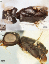
Figure 10. Brightfield image showing the head and mesosoma of Conostigmus bucephalus Mikó and Trietsch sp. nov.
(A) Dorsal view. (B) Lateral view.
Figure 11
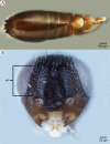
Figure 11. Brightfield image showing the head and metasoma of Conostigmus bucephalus Mikó and Trietsch sp. nov.
(A) Metasoma, dorsal view. (B) Head, anterior view (vrf, ventral region on frons, asr, antennal scrobe).
Figure 12
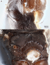
Figure 12. Brightfield image showing the mesosoma of Conostigmus bucephalus Mikó and Trietsch sp. nov.
(A) Pronotum, part of propleuron and part of mesopectus, anterolateral view. (B) Part of mesomoma, posterior view (vrf, ventral region on frons with transverse scutes, asr, antennal scrobe).
Figure 13
Figure 13. Brightfield image showing the head of Conostigmus clavatus Dessart, 1997.
(A) Ventral view. (B) Anterior view (stc, subtorular carina; ita, intertorular area).
Figure 14
Figure 14. Brightfield image showing the head and mesosoma of Conostigmus clavatus Dessart, 1997.
(A) Head, posterior view. (B) Mesosoma, lateral view (poc, preoccipital carina; axc, axillular carina; mfc, median flange of occipital carina; sfc, submedial flange of occipital carina).
Figure 15
Figure 15. Brightfield image showing the head and mesosoma of Conostigmus clavatus Dessart, 1997.
(A) Mesomoma, dorsal view. (B) Head and mesosoma, dorsal view (poc, preoccipital carina; axc, axillular carina; mfc, median flange of occipital carina; sfc, submedial flange of occipital carina).
Figure 16
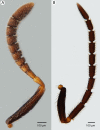
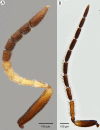
Figure 16. Brightfield image showing the antenna of Conostigmus clavatus Dessart, 1997.
(A) Female. (B) Male.
Figure 17
Figure 17. CLSM volume rendered micrographs showing the male genitalia of Conostigmus clavatus Mikó and Trietsch sp. nov.
(A) Ventral view (B) Dorsal view (ddm, distodorsal margin of cupula; pdc, proximodorsal notch of cupula).
Figure 18
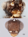
Figure 18. Brightfield image showing the head of Conostigmus madagascariensis Mikó and Trietsch sp. nov.
(A) Head, anterior view. (B) Mandible, labrum and clypeus, anteroventral view.
Figure 19
Figure 19. Brightfield image showing the mesosoma of Conostigmus madagascariensis Mikó and Trietsch sp. nov.
(A) mesosoma, lateral view. (B) Mesosoma, dorsal view (ste, sternaulus).
Figure 20
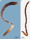
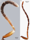
Figure 20. Brightfield image showing the antenna of Conostigmus madagascariensis Mikó and Trietsch sp. nov.
(A) Male. (B) Female.
Figure 21
Figure 21. CLSM volume rendered micrographs showing the male genitalia of Conostigmus fianarantsoaensis Mikó and Trietsch sp. nov.
(A) Ventral view. (B) Dorsal view. (C) Lateroventral view. (D) Dorsal view partially rendered (ppc, proximolateral projection of cupula; pdc, proximodorsal notch of cupula; hrp, harpe; dc, Dorsomedian conjunctiva of the gonostyle/volsella complex; aps, apical parossiculal seta).
Figure 22
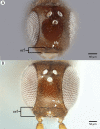
Figure 22. Brightfield image showing the head of Conostigmus longulus Dessart, 1997, anterior view.
(A) Smaller specimen. (B) Larger specimen (tfc, transverse frontal carina; vrf, ventral region of frons; dep, depression surrounding frontal pit).
Figure 23
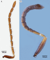
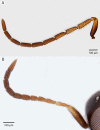
Figure 23. Brightfield image showing the antenna of Conostigmus longulus Dessart, 1997, lateral view.
(A) Female. (B) Male.
Figure 24
Figure 24. Brightfield image showing the head and mesosoma of Conostigmus longulus Dessart, 1997, anterior view
(A) Larger specimen. (B) Smaller specimen.
Figure 25
Figure 25. Brightfield image showing the head and mesosoma of Conostigmus longulus Dessart, 1997, anterior view.
(A) Larger specimen. (B) Smaller specimen.
Figure 26
Figure 26. CLSM volume rendered micrographs showing the male genitalia of Conostigmus longulus Dessart, 1997.
(A) Ventral view. (B) Dorsal view (hrp, harpe; dhs, dorsomedial setae of harpal setal ring).
Figure 27
Figure 27. Brightfield image showing the antenna of Conostigmus lucidus Mikó and Trietsch sp. nov., lateral view.
(A) Female. (B) Male.
Figure 28
Figure 28. Brightfield image showing the head and mesosoma of Conostigmus lucidus Mikó and Trietsch sp. nov.
(A) Head, anterior view. (B) Head and mesosoma, dorsal view (mml, median mesoscutal line).
Figure 29
Figure 29. Brightfield image showing the head and mesosoma of Conostigmus lucidus Mikó and Trietsch sp. nov., lateral view.
(A) Smaller specimen. (B) Larger specimen (ste, sternaulus).
Figure 30
Figure 30. CLSM volume rendered micrographs showing the male genitalia of Conostigmus lucidus Mikó and Trietsch sp. nov.
(A) Ventral view. (B) Dorsal view. (C) Ventral view, partially rendered. (D) Dorsal view, partially rendered. (Arrow on (C) showing bridge connecting parossiculus with gonostyle, on (D) showing lack of dorsomedian conjectiva of gonostyle/volsella complex).
Figure 31
Figure 31. Brightfield image showing the mesosoma of Conostigmus species, posterolateral view.
(A) Conostigmus missyhazenae Mikó and Trietsch sp. nov. (B) Conostigmus lucidus Mikó and Trietsch sp. nov.
Figure 32
Figure 32. Brightfield image showing the head of Conostigmus macrocupula Mikó and Trietsch sp. nov.
(A) Anterior view. (B) Posterior view.
Figure 33
Figure 33. Brightfield image showing the head and antenna of Conostigmus macrocupula Mikó and Trietsch sp. nov.
(A) Head, ventral view. (B) Antenna, lateral view.
Figure 34
Figure 34. Brightfield image showing the head and mesosoma of Conostigmus macrocupula Mikó and Trietsch sp. nov.
(A) Lateral ventral view. (B) Dorsal view.
Figure 35
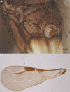
Figure 35. Brightfield image showing the mesosoma and wing of Conostigmus macrocupula Mikó and Trietsch sp. nov.
(A) Mesosoma, posterolateral view. (B) Fore wing.
Figure 36
Figure 36. CLSM volume rendered micrographs showing the male genitalia of Conostigmus macrocupula Mikó and Trietsch sp. nov.
(A) Ventral view. (B) Dorsal view (cup, cupula; pss, parosiculal setae; dc, dorsomedial conjunctiva of gonostyle/volsella complex).
Figure 37
Figure 37. Brightfield image showing the intraspecific variability in mandible structure of Conostigmus madagascariensis Mikó and Trietsch sp. nov.
Figure 38
Figure 38. Brightfield image showing the intraspecific variability in anterior head morphology of Conostigmus madagascariensis Mikó and Trietsch sp. nov.
Figure 39
Figure 39. Brightfield image showing the head of Conostigmus species, posterior view.
(A) Conostigmus madagascariensis Mikó and Trietsch sp. nov. (B) Conostigmus fianarantsoaensis Mikó and Trietsch sp. nov. (pos, preoccipital furrow).
Figure 40
Figure 40. Brightfield image showing the antenna of Conostigmus madagascariensis Mikó and Trietsch sp. nov.
(A) Male. (B) Female.
Figure 41
Figure 41. Brightfield image showing the inraspecific variability of the mesosoma of Conostigmus madagascariensis Mikó and Trietsch sp. nov., dorsal view.
(A) Smaller specimen. (B) Larger specimen (pos, postocellar sulcus).
Figure 42
Figure 42. Brightfield image showing the inraspecific variability of the mesosoma of Conostigmus madagascariensis Mikó and Trietsch sp. nov., lateral view.
(A) Larger specimen. (B) Smaller specimen.
Figure 43
Figure 43. CLSM volume rendered micrographs showing the male genitalia of Conostigmus madagascariensis Mikó and Trietsch sp. nov.
(A) Ventral view. (B) Dorsal view (dc, dorsomedian conjunctiva of gonostyle/volsella complex; hrp, harpe; pdc, proximodorsal notch of cupula; ppc, proximlateral projection of cupula; pss, parossiculal seta).
Figure 44
Figure 44. Brightfield image showing the head of Conostigmus missyhazenae Mikó and Trietsch sp. nov.
(A) Posterior view. (B) Ventral view.
Figure 45
Figure 45. Brightfield image showing the antenna of Conostigmus missyhazenae Mikó and Trietsch sp. nov.
(A) Male. (B) Female.
Figure 46
Figure 46. Brightfield image showing the head and mesosoma of Conostigmus missyhazenae Mikó and Trietsch sp. nov.
(A) Dorsal view. (B) Lateral view.
Figure 47
Figure 47. CLSM volume rendered micrographs showing the male genitalia of Conostigmus missyhazenae Mikó and Trietsch sp. nov.
(A) Ventral view. (B) Dorsal view (hrp, harpe; dhs, dorsomedial setae of harpal setal ring).
Figure 48
Figure 48. Brightfield image showing the head of Conostigmus pseudobabaiax Mikó and Trietsch sp. nov.
(A) Anterior view. (B) Dorsal view.
Figure 49
Figure 49. Brightfield image showing the antenna of Conostigmus pseudobabaiax Mikó and Trietsch sp. nov.
(A) Female. (B) Male.
Figure 50
Figure 50. Brightfield image showing the head and mesosoma of Conostigmus pseudobabaiax Mikó and Trietsch sp. nov.
(A) Mesosoma, dorsal view. (B) Head and mesosoma, lateral view.
Figure 51
Figure 51. Brightfield image showing the middle anteromesoscutum of Conostigmus species, dorsal view.
(A) Conostigmus toliaraensis Mikó and Trietsch sp. nov. (B) Conostigmus pseudobabaiax Mikó and Trietsch sp. nov.
Figure 52
Figure 52. CLSM volume rendered micrographs showing the male genitalia of Conostigmus pseudobabaiax Mikó and Trietsch sp. nov.
(A) Ventral view. (B) Dorsal view (dhs, dorsomedial setae of harpal setal ring; gvs, gonostyle/volsella complex; hrp, harpe; pdc, proximodorsal notch of cupula).
Figure 53
Figure 53. Brightfield image showing the antenna of Conostigmus toliaraensis Mikó and Trietsch sp. nov.
(A) Female. (B) Male.
Figure 54
Figure 54. Brightfield image showing the head of Conostigmus toliaraensis Mikó and Trietsch sp. nov.
(A) Anterior view. (B) Posterior view.
Figure 55
Figure 55. Brightfield image showing the mesosoma of Conostigmus toliaraensis Mikó and Trietsch sp. nov.
Dorsal view (mml, median mesoscutal line).
Figure 56
Figure 56. Brightfield image showing the head and mesosoma of Conostigmus toliaraensis Mikó and Trietsch sp. nov.
(A) Mesosoma, posterior view. (B) Head and mesosoma, lateral view. (ste, sternaulus).
Figure 57
Figure 57. CLSM volume rendered micrographs showing the male genitalia of Conostigmus toliaraensis Mikó and Trietsch sp. nov.
(A) Ventral view (B) Dorsal view (hrp, harpe; dhs, dorsomedial setae of harpal setal ring; gvs, gonostyle/volsella complex; pdc, proximodorsal notch of cupula).
Figure 58
Figure 58. SEM micrograph of the metasoma of Trichosteresis glabra (Boheman, 1831) in ventral view showing scutes corresponding to a possible exocrine gland.

References

    1. Al Khatib F, Fusu L, Cruaud A, Gibson G, Borowiec N, Rasplus J-Y, Ris N, Delvare G. An integrative approach to species discrimination in the Eupelmus urozonus complex (Hymenoptera, Eupelmidae), with the description of 11 new species from the Western Palaearctic. Systematic Entomology. 2014;39(4):806–862. doi: 10.1111/syen.12089. - DOI
    1. Allen RT, Ball GE. Synopsis of Mexican taxa of the Loxandrus series (Coleoptera: Carabidae: Pterostichini) Transactions of the American Entomological Society (1890-) 1979;105(4):481–575.
    1. Araj SA, Wratten SD, Lister AJ, Buckley HL. Floral nectar affects longevity of the aphid parasitoid Aphidius ervi and its hyperparasitoid Dendrocerus aphidum. New Zealand Plant Protection. 2006;59:178.
    1. Azevedo RBR, French V, Partridge L. Temperature modulates epidermal cell size in Drosophila melanogaster. Journal of Insect Physiology. 2002;48(2):231–237. doi: 10.1016/S0022-1910(01)00168-8. - DOI - PubMed
    1. Balhoff JP, Mikó I, Yoder MJ, Mullins PL, Deans AR. A semantic model for species description applied to the ensign wasps (Hymenoptera: Evaniidae) of New Caledonia. Systematic Biology. 2013;62(5):639–659. doi: 10.1093/sysbio/syt028. - DOI - PMC - PubMed

LinkOut - more resources